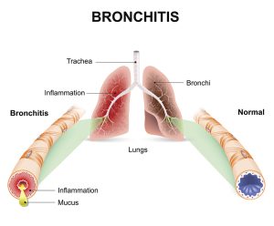
Bronchitis | Causes | Signs and symptoms | Pathophysiology | Treatment | Nursing Management

Two key characteristics of bronchitis include a persistent cough, often producing mucus, and difficulty breathing due to narrowed airways, leading to wheezing and shortness of breath, particularly during physical activity.

Overall, bronchitis affects the respiratory system, particularly the bronchial tubes, and manifests with symptoms such as coughing and breathing difficulties.

Causes of Bronchitis

  • Microorganisms: Bronchitis can be triggered by various microorganisms, including viruses and bacteria. These pathogens infiltrate the bronchial tubes, leading to inflammation and respiratory distress.
  • Extension of Infection from the Trachea: Infections originating in the trachea can extend downward into the bronchial passages, exacerbating inflammation and disrupting normal respiratory function.
  • Traumatic Injuries: Traumatic injuries to the chest area can disrupt the integrity of the respiratory system, potentially leading to inflammation of the bronchial tubes and subsequent bronchitis.
  • Excessive Cold Air: Exposure to excessively cold air can irritate the bronchial tubes, making individuals more susceptible to bronchitis by compromising the respiratory tract’s natural defenses.
  • Smoking: Smoking is a significant risk factor for bronchitis, as the chemicals in tobacco smoke damage the bronchial lining, weaken the immune response, and contribute to chronic inflammation of the airways.
  • Overexposure to Industrial Fumes: Workers exposed to industrial fumes, such as those found in manufacturing or chemical processing environments, are at risk of developing bronchitis due to respiratory irritation and inflammation caused by these harmful substances.

Types

There are 2 types of bronchitis, and these are:

  • Acute bronchitis
  • Chronic bronchitis

Acute bronchitis

Acute bronchitis refers to inflammation of the bronchial tree with minimal structural alterations in the affected areas. It typically presents with symptoms such as coughing or a slight irritation in the throat, sometimes accompanied by the production of sputum.

The primary cause of acute bronchitis is viral infections targeting the bronchial epithelium, leading to inflammation and heightened mucus production within the bronchi.

Causes of Acute Bronchitis

Viral Infections: Acute bronchitis is frequently triggered by viral infections, with several types of viruses implicated in its onset. Common viral culprits include rhinoviruses, coronaviruses, adenoviruses, metapneumoviruses, parainfluenza viruses, respiratory syncytial virus (RSV), and influenza viruses.

These viruses infect the epithelial cells lining the bronchial tubes, provoking inflammation and increased mucus production, which contribute to the characteristic symptoms of acute bronchitis.

Bacterial Infections: While less common than viral causes, bacterial infections can also lead to acute bronchitis. Bacteria such as Mycoplasma pneumoniae, Chlamydophila pneumoniae, Bordetella pertussis, and Streptococcus pneumonia are known to provoke bronchial inflammation and respiratory symptoms.

These bacteria may infect the bronchial epithelium, triggering an immune response and causing the characteristic symptoms of acute bronchitis.

Signs and Symptoms of Acute Bronchitis

  • Dry Cough Progressing to Productive Cough: Initially, acute bronchitis often presents with a dry cough that may later transition to a productive cough, characterized by the expulsion of mucus or phlegm from the bronchial tubes.
  • Chest Pains: Individuals with acute bronchitis may experience discomfort or pain in the chest area, often due to the persistent coughing and inflammation of the bronchial passages.
  • Haemoptysis (Coughing up Blood-Stained Sputum): In some cases, acute bronchitis can lead to haemoptysis, where individuals cough up sputum tinged with blood. This symptom warrants prompt medical attention.
  • General Body Malaise: Acute bronchitis can cause a feeling of overall body malaise, characterized by fatigue, weakness, and a general sense of discomfort or unease.
  • Dyspnoea (Shortness of Breath): Shortness of breath, or dyspnoea, is a common symptom of acute bronchitis, particularly during physical exertion or when breathing becomes more labored due to bronchial inflammation.
  • Fatigue: Fatigue is a prevalent symptom accompanying acute bronchitis, as the body expends energy to combat the underlying infection and cope with the respiratory distress.
  • Sore Throat: Many individuals with acute bronchitis experience a sore throat, which can result from irritation of the throat tissues due to coughing or from the viral or bacterial infection affecting the upper respiratory tract.
  • Cyanosis: In severe cases, cyanosis may occur, characterized by a bluish discoloration of the skin or mucous membranes, indicating a decrease in oxygen saturation in the blood.
  • Nasal Congestion: Acute bronchitis may also be accompanied by nasal congestion, as the respiratory infection can extend to the upper airways, leading to inflammation and congestion of the nasal passages.
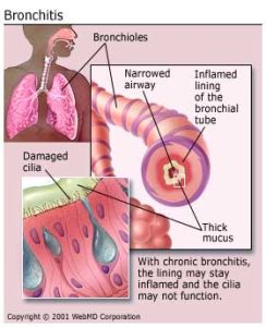
Chronic bronchitis

Chronic bronchitis, categorized as a form of chronic obstructive pulmonary disease (COPD), is characterized by a persistent cough producing mucus for at least three consecutive months per year, spanning a minimum of two years, in the absence of other underlying conditions.

This condition typically arises from repetitive damage to the airways due to the inhalation of irritants. The primary culprit is cigarette smoking, which accounts for the majority of cases, followed by exposure to environmental pollutants like sulfur dioxide or nitrogen dioxide, as well as occupational contact with respiratory irritants.

Individuals who are regularly exposed to cigarette smoke, chemical lung irritants, or who have compromised immune systems face an elevated risk of developing chronic bronchitis (Stanley, 2022).

Causes of Chronic Bronchitis

Chronic bronchitis predominantly arises from several key factors:

  • Cigarette Smoking and Tobacco Use: The leading cause of chronic bronchitis is cigarette smoking or the use of other tobacco products. The inhalation of tobacco smoke damages the bronchial tubes over time, leading to chronic inflammation and mucus production.
  • Chronic Inhalation of Air Pollution: Prolonged exposure to environmental pollutants, including airborne particulate matter and toxic fumes, contributes significantly to the development of chronic bronchitis. Individuals living in areas with high levels of air pollution are particularly at risk.
  • Occupational Exposures: Certain occupations involving exposure to hazardous substances pose a heightened risk of chronic bronchitis. Workers in industries such as coal mining, grain handling, textile manufacturing, livestock farming, and metal molding are frequently exposed to irritating fumes, dust, or chemicals, which can cause chronic inflammation of the bronchial passages.
  • Coal Mining: Workers in coal mining environments are exposed to coal dust and other airborne particles, which can irritate the respiratory tract and contribute to the development of chronic bronchitis over time.
  • Grain Handling: Grain handlers are exposed to grain dust, which contains various allergens and irritants that can exacerbate respiratory conditions like chronic bronchitis with prolonged exposure.
  • Textile Manufacturing: Workers in textile manufacturing facilities may inhale fibers, dyes, and chemicals present in the air, leading to chronic irritation of the bronchial tubes and increasing the risk of developing chronic bronchitis.
  • Livestock Farming: Individuals involved in livestock farming, such as farmers and agricultural workers, are exposed to organic dust, animal dander, and airborne pathogens, which can contribute to chronic bronchitis development.
  • Metal Molding: Workers in metal molding industries are exposed to metal fumes and dust, which can irritate the airways and contribute to the onset of chronic bronchitis.

Pathophysiology of Chronic Bronchitis

Chronic bronchitis is characterized by a series of pathological changes in the bronchial tubes due to prolonged exposure to irritants. This exposure triggers hypertrophy and hyperplasia of the bronchial mucous glands, leading to an increase in mucus production.

Damage to the cilia, which normally sweep away dust, irritants, and mucus, occurs alongside squamous metaplasia of the columnar epithelium. Chronic inflammation with an infiltration of leukocytes and lymphocytes into the bronchial walls further exacerbates the condition.

Excessive secretion from goblet cells obstructs the movement of cilia, impairing their ability to clear the airway effectively. This loss of cilia alters the function of alveolar macrophages, increasing their susceptibility to bronchial infections.

Consequently, the airway becomes obstructed, with mucus and debris accumulating and narrowing the airway lumen, leading to decreased airflow. This increased resistance to airflow imposes greater work on breathing.

Continued inflammation results in ulceration of the epithelial layer. Upon healing, the walls of the airways thicken, further narrowing the passages.

This progressive narrowing of the airways contributes to the hallmark symptoms of chronic bronchitis, including persistent coughing, increased mucus production, and difficulty breathing.

Signs and Symptoms of Chronic Bronchitis

Chronic bronchitis presents with a range of persistent symptoms indicative of ongoing airway inflammation and mucus production:

  • Chronic Cough: A hallmark symptom of chronic bronchitis is a persistent cough, resulting from repeated irritation of the bronchial tubes over time.
  • Copious, Thick, and Brown Sputum: Individuals with chronic bronchitis often produce abundant, thick, and brown-colored sputum, reflecting the increased secretion of mucus from goblet cells in the bronchial lining.
  • Haemoptysis: Haemoptysis, or coughing up blood, may occur in certain cases of chronic bronchitis, indicating bleeding from specific areas of the bronchial tree affected by inflammation and irritation.
  • Dyspnoea: Shortness of breath, or dyspnoea, is common in chronic bronchitis due to the constriction of the bronchioles caused by inflammation and the presence of excessive mucus, impeding airflow.
  • Chest Pains: Chest pains may arise due to reduced airspaces within the lungs, resulting from chronic inflammation and mucus accumulation, leading to feelings of tightness or discomfort in the chest.
  • Cyanosis: Cyanosis, characterized by a bluish discoloration of the skin or mucous membranes, can occur due to insufficient oxygenation of the blood (hypoxia) resulting from impaired lung function in chronic bronchitis.
  • Fever: In some cases, fever may accompany chronic bronchitis, particularly during exacerbations or in response to secondary respiratory infections.
  • Wheezing: Wheezing, a high-pitched whistling sound during breathing, may occur due to the accumulation of mucus in the narrowed airways, leading to turbulent airflow and respiratory distress.

Medical Management

History and Physical Examination for Bronchitis:

History

  • Symptom Duration and Severity: Ask the patient about the duration and severity of symptoms such as cough, sputum production, dyspnea, chest pain, and fatigue.
  • Risk Factors: Ask about patient risk factors such as smoking history, occupational exposures, environmental pollution, and previous respiratory infections.
  • Medical History: Ask for any pre-existing conditions such as asthma, allergies, COPD, or immunocompromised status.
  • Recent Illnesses: Ask if the patient has had recent upper respiratory infections or exacerbations of chronic respiratory conditions.
  • Medication History: Assess for any recent medication use, including antibiotics, bronchodilators, or corticosteroids.
  • Functional Impact: Evaluate how symptoms affect daily activities, sleep, and overall quality of life.

Physical Examination

  • Vital Signs: Measure vital signs, including temperature, heart rate, respiratory rate, and oxygen saturation.
  • Respiratory Examination: Auscultate lung fields for abnormal breath sounds such as wheezes, crackles, or diminished breath sounds. Evaluate respiratory effort and chest expansion.
  • Cough Assessment: Observe the character of the cough (dry or productive), and inquire about factors that exacerbate or alleviate coughing.
  • Sputum Evaluation: Assess the quantity, color, and consistency of the sputum produced.
  • Chest Examination: Palpate the chest for tenderness or deformities. Percuss to assess for dullness or hyper-resonance, indicating possible consolidation or hyperinflation.
  • Cardiovascular Examination: Perform a focused cardiovascular examination to evaluate for signs of cardiac involvement or complications.
  • General Examination: Assess for signs of systemic illness such as fever, malaise, weight loss, or peripheral edema.

Treatment for Bronchitis

Effective management of bronchitis involves a multifaceted approach aimed at relieving symptoms, reducing inflammation, and addressing the underlying cause:

  • Antibiotics: If the bronchitis is caused by a bacterial infection, antibiotics such as ampicillin may be prescribed to target the bacterial pathogens and prevent complications. However, antibiotics are not routinely recommended for viral bronchitis.
  • Anti-Inflammatory Drugs: Anti-inflammatory medications like prednisolone may be prescribed to reduce inflammation in the airways, alleviate swelling, and ease breathing difficulties associated with bronchitis.
  • Cough expectorants: Cough expectorants such as ammonium chloride may be used to help thin and loosen mucus, making it easier to cough up and expel from the airways. This can help relieve coughs and promote the clearance of respiratory secretions.
  • Steam Inhalations: Steam inhalations can help moisten and loosen mucus, making it easier to clear from the airways. Inhaling steam from a bowl of hot water or using a humidifier can provide relief from congestion and soothe irritated airways.
  • Environmental Considerations: Creating a warm, well-ventilated room environment can help ease symptoms of bronchitis. Avoiding exposure to cold air, tobacco smoke, and other respiratory irritants can help prevent the exacerbation of symptoms.
  • Hydration and Nutrition: Maintaining hydration is important during bronchitis to help thin mucus and prevent dehydration. Drinking plenty of fluids and consuming a light, nutritious diet can support the immune system and aid in recovery.
  • Rest and Comfort Measures: Encouraging adequate rest and relaxation can support the body’s healing process during bronchitis. Providing comfort measures such as warm blankets and soothing throat lozenges can help alleviate discomfort and promote recovery.
  • Monitoring and Follow-Up: Regular monitoring of symptoms and follow-up with healthcare providers is important to track progress and ensure appropriate management of bronchitis. In severe cases or if symptoms persist or worsen, further medical evaluation may be necessary.

Investigations for Bronchitis

Effective diagnosis of bronchitis involves a comprehensive assessment combining various investigative techniques:

  • Chest X-ray: Chest X-rays may reveal hyperinfiltration and increased bronchovascular markings, providing insights into the extent of inflammation and potential complications such as pneumonia or chronic obstructive pulmonary disease (COPD).
  • Pulmonary Function Tests (PFTs): Pulmonary function tests, including spirometry, assess lung function parameters such as vital capacity, forced expiratory flow, and residual volume. In bronchitis, PFTs typically demonstrate increased residual volume, decreased vital capacity, and forced expiratory flow, along with normal static compliance and diffusion capacity.
  • Arterial Blood Gas (ABG) Analysis: Arterial blood gas analysis provides valuable information about oxygenation and ventilation status. In bronchitis, ABG analysis often reveals decreased arterial oxygen tension (PaO2) due to impaired gas exchange, along with normal or increased arterial carbon dioxide tension (PaCO2), indicating respiratory compensation.
  • Sputum Culture: Sputum culture may be performed to identify the presence of microorganisms and assess neutrophil levels, aiding in determining the underlying cause of bronchitis and guiding appropriate antibiotic therapy if bacterial infection is suspected.
  • Electrocardiogram (ECG): An electrocardiogram may be conducted to assess cardiac function, particularly in cases where bronchitis coexists with cardiovascular conditions. ECG findings may reveal atrial arrhythmias, which can occur secondary to hypoxemia or as a consequence of chronic lung disease.

Nursing Management

Aims

  • To maintain clear airway
  • To maintain circulation
  • To promote quick recovery
  • To prevent complications such as respiratory failure

Optimal Patient Environment

  • Nurse the patient in a well-ventilated room situated conveniently close to the nurse’s station for easy monitoring.
  • Equip the room with all essential medical supplies and bed accessories, including an oxygen cylinder, suctioning machine, drip stand, and adjustable backrest, ensuring prompt access to necessary equipment.
  • Maintain a serene and tranquil environment conducive to rest and recovery, minimizing noise to facilitate uninterrupted rest periods.
  • Enhance humidity levels in the room by utilizing steam generated from boiling water, aiding in the liquefaction of respiratory secretions, and promoting airway clearance.

Optimal Positioning

  • Position the patient comfortably in the semi-Fowler’s position with the assistance of a backrest and strategically placed pillows to facilitate optimal lung expansion and alleviate dyspnea.
  • The semi-Fowler’s position involves reclining the patient’s upper body at approximately a 45-degree angle, which helps reduce the workload on the respiratory muscles and promotes more efficient breathing.
  • By elevating the head and upper body, this position encourages the expansion of the lungs and allows for better ventilation of the alveoli.
  • Additionally, using pillows to support the back and provide cushioning under the head and neck can enhance patient comfort and stability in this position, minimizing the risk of discomfort or pressure sores.
  • Overall, positioning the patient in the semi-Fowler’s position optimizes respiratory function while promoting comfort and relaxation during the recovery process.

Psychological Support

  • Give clear explanations of the disease process to both the patient and their family members to alleviate anxiety and promote understanding.
  • Encourage open communication by addressing any questions or concerns the patient or family may have, fostering a supportive environment where they feel comfortable expressing their feelings and uncertainties about the illness.
  • Actively involve the patient and their family in care decisions, empowering them to participate in their treatment plan and providing opportunities for shared decision-making.
  • Offer referrals to additional support services as needed to further address emotional and psychological needs.
  • When introducing medical equipment, such as oxygen therapy, take the time to explain its purpose and function to the patient, reassuring them and easing any anxieties they may have.
  • Similarly, provide thorough explanations of all procedures performed on the patient, outlining the steps involved and the intended outcomes, to enhance cooperation and promote patient engagement in their care.
  • By prioritizing patient education and communication, healthcare providers can help alleviate anxiety, build trust, and foster a collaborative approach to managing the patient’s condition.

Patient Observation

  • Regularly assess vital signs, including temperature, pulse, respiration, and blood pressure, to track the patient’s physiological status and identify any deviations from baseline values.
  • Monitor sputum characteristics such as consistency, quality, amount, and color to assess respiratory secretions and detect changes indicative of infection or inflammation.
  • Observe the patient’s breathing pattern for signs of dyspnea, wheezing, or other respiratory distress symptoms. Continuously evaluate the patient’s response to treatment, noting any improvements or adverse reactions to medications.
  • Monitor for potential side effects of drugs and promptly address any concerns or complications that may arise.
  • Assess changes in respiratory function through objective measures such as pulmonary function tests or arterial blood gas analysis.
  • Additionally, weigh the patient three times weekly and assess for signs of edema to identify potential complications such as cor pulmonale or fluid overload.
  • Regular and thorough observation allows healthcare providers to intervene promptly and adjust treatment strategies as needed to optimize patient outcomes.

Nutritional Support

  • Offer the patient a diet rich in high-calorie, protein-packed foods to support the healing process and promote recovery.
  • Give a patient small, frequent meals to help conserve the patient’s energy levels and prevent fatigue.
  • Ensure adequate hydration by encouraging the intake of at least 3 liters of fluids daily, which helps to loosen respiratory secretions.
  • Schedule respiratory therapy sessions at least 1 hour before or after meals to minimize any potential discomfort or interference with digestion.
  • Additionally, provide thorough mouth care following bronchodilator inhalation therapy to maintain oral hygiene and prevent complications.
  • By prioritizing nutritional support and hydration, healthcare providers can contribute to the patient’s overall well-being and optimize their response to treatment.

Monitoring Elimination

  • Regularly monitor and document both fluid intake and output to assess renal function and ensure proper kidney function.
  • Give a patient a diet rich in roughage to prevent constipation, thereby reducing the strain on the heart.
  • By promoting regular bowel movements and supporting renal health, these measures contribute to the overall well-being and comfort of the patient.
  • Give the patient ample fluids throughout the day to promote hydration and support renal function. Offer water, herbal teas, and hydrating beverages to ensure adequate fluid intake.
  • Proper hydration helps maintain optimal renal blood flow and promotes the excretion of waste products, supporting renal function and preventing complications such as dehydration or urinary tract infections.
  • Develop and implement a bowel management plan that includes dietary modifications, regular toileting schedules, and the use of stool softeners or laxatives as needed to prevent constipation.
  • Incorporate high-fiber foods such as fruits, vegetables, and whole grains into the patient’s diet to promote regular bowel movements and reduce the risk of straining.
  • Preventing constipation reduces the risk of complications such as fecal impaction or increased intra-abdominal pressure, which can negatively impact renal function and exacerbate cardiovascular strain.

Hygiene

  • During the acute phase, administer bedside baths to the patient to enhance blood circulation and bolster self-esteem. Encourage oral care to stimulate salivation and maintain oral hygiene.
  • Ensure the bed is properly made, and change soiled linens promptly to optimize comfort. Offer a clean sputum mug with a lid to minimize the risk of infection spreading.
  • Provide a designated bin for the disposal of tissues to prevent the transmission of infection.
  • Emphasize the importance of handwashing after handling secretions to minimize the risk of contamination and infection transmission.
  • These hygiene measures are essential for promoting patient comfort and reducing the spread of pathogens in the healthcare environment.

Rest and Exercise

  • Establish a tranquil environment conducive to rest, facilitating the patient’s recovery process.
  • Administer chest physiotherapy techniques, such as postural drainage and chest percussion, to optimize respiratory function and promote airway clearance.
  • Assist the patient in alternating between periods of rest and gentle activity to conserve energy levels and minimize fatigue.
  • Encourage engagement in daily activities and provide appropriate diversional activities to enhance overall well-being.
  • These interventions support a balanced approach to rest and exercise, contributing to the patient’s physical and psychological recovery.

Medication

  • Ensure timely administration of prescribed medications and monitor the patient’s response to treatment closely.
  • Encourage the patient to take prescribed medications as per schedule to promote a quick recovery.
  • Teach the patient about the side effects of these drugs in order to raise awareness.
  • Encourage the patient to report any side effects as soon as possible to the hospital for prompt management.
  • Instruct the patient to stop the drug from losing its potency.

Information and Education

  • Advise the patient to steer clear of crowded places and individuals with known infections, and ensure they receive influenza and pneumococcus vaccinations.
  • Educate both the patient and their family on performing postural drainage and chest percussion. Guide them to maintain each position for 10 minutes before proceeding with percussion, followed by coughing.
  • Additionally, instruct on coughing and deep-breathing techniques to enhance ventilation and facilitate secretion removal.
  • Review all prescribed medications, including dosage, potential adverse effects, and intended purposes. Encourage the patient to promptly report any adverse reactions to their physician.
  • Stress the importance of consuming high-calorie, protein-rich meals and staying well-hydrated to prevent dehydration and aid in secretion clearance.
  • If the patient is a smoker, strongly advise them to quit smoking. Offer smoking cessation counseling or resources if needed.
  • Encourage the avoidance of irritants such as automobile exhaust fumes, aerosol sprays, and industrial pollutants to minimize respiratory irritation.
  • Warn against exposure to cold air blasts, which can trigger bronchospasm. Recommend avoiding cold, windy weather or using a scarf or mask to cover the mouth and nose when outdoors.

Complications of Bronchitis

  • Bronchopneumonia: Inflammation and infection of the lung tissue can lead to bronchopneumonia, a serious complication characterized by the consolidation of lung tissue and impaired gas exchange.
  • Recurrent Lower Respiratory Tract Infections: Chronic bronchitis predisposes individuals to repeated infections of the lower respiratory tract, which can exacerbate symptoms and contribute to respiratory compromise over time.
  • Cor Pulmonale: Chronic obstruction in the lungs can result in cor pulmonale, a condition marked by right-sided heart failure due to increased pressure in the pulmonary circulation, often leading to hypertrophy and dilation of the right ventricle.
  • Acute Respiratory Failure: Severe bronchitis can progress to acute respiratory failure, a life-threatening condition characterized by inadequate gas exchange, hypoxemia, and hypercapnia, requiring immediate medical intervention and respiratory support.
  • Pneumonia: Bronchitis can predispose individuals to pneumonia, a potentially severe infection of the lung parenchyma often caused by bacterial or viral pathogens spreading from the bronchial tubes to the lung tissue.
  • Right-Sided Heart Failure: Increased pressure on the right side of the heart to pump blood into the lungs against resistance can lead to right-sided heart failure, resulting in systemic congestion and fluid retention.
  • Atelectasis and Bronchiectasis: Chronic bronchitis can contribute to the development of complications such as atelectasis, the collapse of lung tissue, and bronchiectasis, the chronic dilation and destruction of bronchial walls, further compromising respiratory function and increasing susceptibility to infections.

Read more: Medical-Surgical Nursing

Read more: Asthma | Classifications | Risk Factors | Sign and symptoms | Pathophysiology | Nursing Management

Facebook
Twitter
LinkedIn
WhatsApp

Recent Posts

Contact Form