Bronchitis | Causes | Signs and symptoms | Pathophysiology | Treatment | Nursing Management

What is bronchitis?

Bronchitis is a respiratory condition characterized by inflammation of the bronchial tubes, the air passages that carry air to the lungs, commonly caused by viral or bacterial infections or exposure to irritants like tobacco smoke or air pollution.
Two key characteristics of bronchitis include a persistent cough, often producing mucus, and difficulty breathing due to narrowed airways, leading to wheezing and shortness of breath, particularly during physical activity.

Overall, bronchitis affects the respiratory system, particularly the bronchial tubes, and manifests with symptoms such as coughing and breathing difficulties.

Causes of Bronchitis

  • Microorganisms: Bronchitis can be triggered by various microorganisms, including viruses and bacteria. These pathogens infiltrate the bronchial tubes, leading to inflammation and respiratory distress.
  • Extension of Infection from the Trachea: Infections originating in the trachea can extend downward into the bronchial passages, exacerbating inflammation and disrupting normal respiratory function.
  • Traumatic Injuries: Traumatic injuries to the chest area can disrupt the integrity of the respiratory system, potentially leading to inflammation of the bronchial tubes and subsequent bronchitis.
  • Excessive Cold Air: Exposure to excessively cold air can irritate the bronchial tubes, making individuals more susceptible to bronchitis by compromising the respiratory tract’s natural defenses.
  • Smoking: Smoking is a significant risk factor for bronchitis, as the chemicals in tobacco smoke damage the bronchial lining, weaken the immune response, and contribute to chronic inflammation of the airways.
  • Overexposure to Industrial Fumes: Workers exposed to industrial fumes, such as those found in manufacturing or chemical processing environments, are at risk of developing bronchitis due to respiratory irritation and inflammation caused by these harmful substances.

Types

There are 2 types of bronchitis, and these are:

  • Acute bronchitis
  • Chronic bronchitis

Acute bronchitis

Acute bronchitis refers to inflammation of the bronchial tree with minimal structural alterations in the affected areas. It typically presents with symptoms such as coughing or a slight irritation in the throat, sometimes accompanied by the production of sputum.

The primary cause of acute bronchitis is viral infections targeting the bronchial epithelium, leading to inflammation and heightened mucus production within the bronchi.

Causes of Acute Bronchitis

Viral Infections: Acute bronchitis is frequently triggered by viral infections, with several types of viruses implicated in its onset. Common viral culprits include rhinoviruses, coronaviruses, adenoviruses, metapneumoviruses, parainfluenza viruses, respiratory syncytial virus (RSV), and influenza viruses.

These viruses infect the epithelial cells lining the bronchial tubes, provoking inflammation and increased mucus production, which contribute to the characteristic symptoms of acute bronchitis.

Bacterial Infections: While less common than viral causes, bacterial infections can also lead to acute bronchitis. Bacteria such as Mycoplasma pneumoniae, Chlamydophila pneumoniae, Bordetella pertussis, and Streptococcus pneumonia are known to provoke bronchial inflammation and respiratory symptoms.

These bacteria may infect the bronchial epithelium, triggering an immune response and causing the characteristic symptoms of acute bronchitis.

Signs and Symptoms of Acute Bronchitis

  • Dry Cough Progressing to Productive Cough: Initially, acute bronchitis often presents with a dry cough that may later transition to a productive cough, characterized by the expulsion of mucus or phlegm from the bronchial tubes.
  • Chest Pains: Individuals with acute bronchitis may experience discomfort or pain in the chest area, often due to the persistent coughing and inflammation of the bronchial passages.
  • Haemoptysis (Coughing up Blood-Stained Sputum): In some cases, acute bronchitis can lead to haemoptysis, where individuals cough up sputum tinged with blood. This symptom warrants prompt medical attention.
  • General Body Malaise: Acute bronchitis can cause a feeling of overall body malaise, characterized by fatigue, weakness, and a general sense of discomfort or unease.
  • Dyspnoea (Shortness of Breath): Shortness of breath, or dyspnoea, is a common symptom of acute bronchitis, particularly during physical exertion or when breathing becomes more labored due to bronchial inflammation.
  • Fatigue: Fatigue is a prevalent symptom accompanying acute bronchitis, as the body expends energy to combat the underlying infection and cope with the respiratory distress.
  • Sore Throat: Many individuals with acute bronchitis experience a sore throat, which can result from irritation of the throat tissues due to coughing or from the viral or bacterial infection affecting the upper respiratory tract.
  • Cyanosis: In severe cases, cyanosis may occur, characterized by a bluish discoloration of the skin or mucous membranes, indicating a decrease in oxygen saturation in the blood.
  • Nasal Congestion: Acute bronchitis may also be accompanied by nasal congestion, as the respiratory infection can extend to the upper airways, leading to inflammation and congestion of the nasal passages.

Chronic bronchitis

Chronic bronchitis, categorized as a form of chronic obstructive pulmonary disease (COPD), is characterized by a persistent cough producing mucus for at least three consecutive months per year, spanning a minimum of two years, in the absence of other underlying conditions.

This condition typically arises from repetitive damage to the airways due to the inhalation of irritants. The primary culprit is cigarette smoking, which accounts for the majority of cases, followed by exposure to environmental pollutants like sulfur dioxide or nitrogen dioxide, as well as occupational contact with respiratory irritants.

Individuals who are regularly exposed to cigarette smoke, chemical lung irritants, or who have compromised immune systems face an elevated risk of developing chronic bronchitis (Stanley, 2022).

Causes of Chronic Bronchitis

Chronic bronchitis predominantly arises from several key factors:

  • Cigarette Smoking and Tobacco Use: The leading cause of chronic bronchitis is cigarette smoking or the use of other tobacco products. The inhalation of tobacco smoke damages the bronchial tubes over time, leading to chronic inflammation and mucus production.
  • Chronic Inhalation of Air Pollution: Prolonged exposure to environmental pollutants, including airborne particulate matter and toxic fumes, contributes significantly to the development of chronic bronchitis. Individuals living in areas with high levels of air pollution are particularly at risk.
  • Occupational Exposures: Certain occupations involving exposure to hazardous substances pose a heightened risk of chronic bronchitis. Workers in industries such as coal mining, grain handling, textile manufacturing, livestock farming, and metal molding are frequently exposed to irritating fumes, dust, or chemicals, which can cause chronic inflammation of the bronchial passages.
  • Coal Mining: Workers in coal mining environments are exposed to coal dust and other airborne particles, which can irritate the respiratory tract and contribute to the development of chronic bronchitis over time.
  • Grain Handling: Grain handlers are exposed to grain dust, which contains various allergens and irritants that can exacerbate respiratory conditions like chronic bronchitis with prolonged exposure.
  • Textile Manufacturing: Workers in textile manufacturing facilities may inhale fibers, dyes, and chemicals present in the air, leading to chronic irritation of the bronchial tubes and increasing the risk of developing chronic bronchitis.
  • Livestock Farming: Individuals involved in livestock farming, such as farmers and agricultural workers, are exposed to organic dust, animal dander, and airborne pathogens, which can contribute to chronic bronchitis development.
  • Metal Molding: Workers in metal molding industries are exposed to metal fumes and dust, which can irritate the airways and contribute to the onset of chronic bronchitis.

Pathophysiology of Chronic Bronchitis

Chronic bronchitis is characterized by a series of pathological changes in the bronchial tubes due to prolonged exposure to irritants. This exposure triggers hypertrophy and hyperplasia of the bronchial mucous glands, leading to an increase in mucus production.

Damage to the cilia, which normally sweep away dust, irritants, and mucus, occurs alongside squamous metaplasia of the columnar epithelium. Chronic inflammation with an infiltration of leukocytes and lymphocytes into the bronchial walls further exacerbates the condition.

Excessive secretion from goblet cells obstructs the movement of cilia, impairing their ability to clear the airway effectively. This loss of cilia alters the function of alveolar macrophages, increasing their susceptibility to bronchial infections.

Consequently, the airway becomes obstructed, with mucus and debris accumulating and narrowing the airway lumen, leading to decreased airflow. This increased resistance to airflow imposes greater work on breathing.

Continued inflammation results in ulceration of the epithelial layer. Upon healing, the walls of the airways thicken, further narrowing the passages.

This progressive narrowing of the airways contributes to the hallmark symptoms of chronic bronchitis, including persistent coughing, increased mucus production, and difficulty breathing.

Signs and Symptoms of Chronic Bronchitis

Chronic bronchitis presents with a range of persistent symptoms indicative of ongoing airway inflammation and mucus production:

  • Chronic Cough: A hallmark symptom of chronic bronchitis is a persistent cough, resulting from repeated irritation of the bronchial tubes over time.
  • Copious, Thick, and Brown Sputum: Individuals with chronic bronchitis often produce abundant, thick, and brown-colored sputum, reflecting the increased secretion of mucus from goblet cells in the bronchial lining.
  • Haemoptysis: Haemoptysis, or coughing up blood, may occur in certain cases of chronic bronchitis, indicating bleeding from specific areas of the bronchial tree affected by inflammation and irritation.
  • Dyspnoea: Shortness of breath, or dyspnoea, is common in chronic bronchitis due to the constriction of the bronchioles caused by inflammation and the presence of excessive mucus, impeding airflow.
  • Chest Pains: Chest pains may arise due to reduced airspaces within the lungs, resulting from chronic inflammation and mucus accumulation, leading to feelings of tightness or discomfort in the chest.
  • Cyanosis: Cyanosis, characterized by a bluish discoloration of the skin or mucous membranes, can occur due to insufficient oxygenation of the blood (hypoxia) resulting from impaired lung function in chronic bronchitis.
  • Fever: In some cases, fever may accompany chronic bronchitis, particularly during exacerbations or in response to secondary respiratory infections.
  • Wheezing: Wheezing, a high-pitched whistling sound during breathing, may occur due to the accumulation of mucus in the narrowed airways, leading to turbulent airflow and respiratory distress.

Medical Management

History and Physical Examination for Bronchitis:

History

  • Symptom Duration and Severity: Ask the patient about the duration and severity of symptoms such as cough, sputum production, dyspnea, chest pain, and fatigue.
  • Risk Factors: Ask about patient risk factors such as smoking history, occupational exposures, environmental pollution, and previous respiratory infections.
  • Medical History: Ask for any pre-existing conditions such as asthma, allergies, COPD, or immunocompromised status.
  • Recent Illnesses: Ask if the patient has had recent upper respiratory infections or exacerbations of chronic respiratory conditions.
  • Medication History: Assess for any recent medication use, including antibiotics, bronchodilators, or corticosteroids.
  • Functional Impact: Evaluate how symptoms affect daily activities, sleep, and overall quality of life.

Physical Examination

  • Vital Signs: Measure vital signs, including temperature, heart rate, respiratory rate, and oxygen saturation.
  • Respiratory Examination: Auscultate lung fields for abnormal breath sounds such as wheezes, crackles, or diminished breath sounds. Evaluate respiratory effort and chest expansion.
  • Cough Assessment: Observe the character of the cough (dry or productive), and inquire about factors that exacerbate or alleviate coughing.
  • Sputum Evaluation: Assess the quantity, color, and consistency of the sputum produced.
  • Chest Examination: Palpate the chest for tenderness or deformities. Percuss to assess for dullness or hyper-resonance, indicating possible consolidation or hyperinflation.
  • Cardiovascular Examination: Perform a focused cardiovascular examination to evaluate for signs of cardiac involvement or complications.
  • General Examination: Assess for signs of systemic illness such as fever, malaise, weight loss, or peripheral edema.

Treatment for Bronchitis

Effective management of bronchitis involves a multifaceted approach aimed at relieving symptoms, reducing inflammation, and addressing the underlying cause:

  • Antibiotics: If the bronchitis is caused by a bacterial infection, antibiotics such as ampicillin may be prescribed to target the bacterial pathogens and prevent complications. However, antibiotics are not routinely recommended for viral bronchitis.
  • Anti-Inflammatory Drugs: Anti-inflammatory medications like prednisolone may be prescribed to reduce inflammation in the airways, alleviate swelling, and ease breathing difficulties associated with bronchitis.
  • Cough expectorants: Cough expectorants such as ammonium chloride may be used to help thin and loosen mucus, making it easier to cough up and expel from the airways. This can help relieve coughs and promote the clearance of respiratory secretions.
  • Steam Inhalations: Steam inhalations can help moisten and loosen mucus, making it easier to clear from the airways. Inhaling steam from a bowl of hot water or using a humidifier can provide relief from congestion and soothe irritated airways.
  • Environmental Considerations: Creating a warm, well-ventilated room environment can help ease symptoms of bronchitis. Avoiding exposure to cold air, tobacco smoke, and other respiratory irritants can help prevent the exacerbation of symptoms.
  • Hydration and Nutrition: Maintaining hydration is important during bronchitis to help thin mucus and prevent dehydration. Drinking plenty of fluids and consuming a light, nutritious diet can support the immune system and aid in recovery.
  • Rest and Comfort Measures: Encouraging adequate rest and relaxation can support the body’s healing process during bronchitis. Providing comfort measures such as warm blankets and soothing throat lozenges can help alleviate discomfort and promote recovery.
  • Monitoring and Follow-Up: Regular monitoring of symptoms and follow-up with healthcare providers is important to track progress and ensure appropriate management of bronchitis. In severe cases or if symptoms persist or worsen, further medical evaluation may be necessary.

Investigations for Bronchitis

Effective diagnosis of bronchitis involves a comprehensive assessment combining various investigative techniques:

  • Chest X-ray: Chest X-rays may reveal hyperinfiltration and increased bronchovascular markings, providing insights into the extent of inflammation and potential complications such as pneumonia or chronic obstructive pulmonary disease (COPD).
  • Pulmonary Function Tests (PFTs): Pulmonary function tests, including spirometry, assess lung function parameters such as vital capacity, forced expiratory flow, and residual volume. In bronchitis, PFTs typically demonstrate increased residual volume, decreased vital capacity, and forced expiratory flow, along with normal static compliance and diffusion capacity.
  • Arterial Blood Gas (ABG) Analysis: Arterial blood gas analysis provides valuable information about oxygenation and ventilation status. In bronchitis, ABG analysis often reveals decreased arterial oxygen tension (PaO2) due to impaired gas exchange, along with normal or increased arterial carbon dioxide tension (PaCO2), indicating respiratory compensation.
  • Sputum Culture: Sputum culture may be performed to identify the presence of microorganisms and assess neutrophil levels, aiding in determining the underlying cause of bronchitis and guiding appropriate antibiotic therapy if bacterial infection is suspected.
  • Electrocardiogram (ECG): An electrocardiogram may be conducted to assess cardiac function, particularly in cases where bronchitis coexists with cardiovascular conditions. ECG findings may reveal atrial arrhythmias, which can occur secondary to hypoxemia or as a consequence of chronic lung disease.

Nursing Management

Aims

  • To maintain clear airway
  • To maintain circulation
  • To promote quick recovery
  • To prevent complications such as respiratory failure

Optimal Patient Environment

  • Nurse the patient in a well-ventilated room situated conveniently close to the nurse’s station for easy monitoring.
  • Equip the room with all essential medical supplies and bed accessories, including an oxygen cylinder, suctioning machine, drip stand, and adjustable backrest, ensuring prompt access to necessary equipment.
  • Maintain a serene and tranquil environment conducive to rest and recovery, minimizing noise to facilitate uninterrupted rest periods.
  • Enhance humidity levels in the room by utilizing steam generated from boiling water, aiding in the liquefaction of respiratory secretions, and promoting airway clearance.

Optimal Positioning

  • Position the patient comfortably in the semi-Fowler’s position with the assistance of a backrest and strategically placed pillows to facilitate optimal lung expansion and alleviate dyspnea.
  • The semi-Fowler’s position involves reclining the patient’s upper body at approximately a 45-degree angle, which helps reduce the workload on the respiratory muscles and promotes more efficient breathing.
  • By elevating the head and upper body, this position encourages the expansion of the lungs and allows for better ventilation of the alveoli.
  • Additionally, using pillows to support the back and provide cushioning under the head and neck can enhance patient comfort and stability in this position, minimizing the risk of discomfort or pressure sores.
  • Overall, positioning the patient in the semi-Fowler’s position optimizes respiratory function while promoting comfort and relaxation during the recovery process.

Psychological Support

  • Give clear explanations of the disease process to both the patient and their family members to alleviate anxiety and promote understanding.
  • Encourage open communication by addressing any questions or concerns the patient or family may have, fostering a supportive environment where they feel comfortable expressing their feelings and uncertainties about the illness.
  • Actively involve the patient and their family in care decisions, empowering them to participate in their treatment plan and providing opportunities for shared decision-making.
  • Offer referrals to additional support services as needed to further address emotional and psychological needs.
  • When introducing medical equipment, such as oxygen therapy, take the time to explain its purpose and function to the patient, reassuring them and easing any anxieties they may have.
  • Similarly, provide thorough explanations of all procedures performed on the patient, outlining the steps involved and the intended outcomes, to enhance cooperation and promote patient engagement in their care.
  • By prioritizing patient education and communication, healthcare providers can help alleviate anxiety, build trust, and foster a collaborative approach to managing the patient’s condition.

Patient Observation

  • Regularly assess vital signs, including temperature, pulse, respiration, and blood pressure, to track the patient’s physiological status and identify any deviations from baseline values.
  • Monitor sputum characteristics such as consistency, quality, amount, and color to assess respiratory secretions and detect changes indicative of infection or inflammation.
  • Observe the patient’s breathing pattern for signs of dyspnea, wheezing, or other respiratory distress symptoms. Continuously evaluate the patient’s response to treatment, noting any improvements or adverse reactions to medications.
  • Monitor for potential side effects of drugs and promptly address any concerns or complications that may arise.
  • Assess changes in respiratory function through objective measures such as pulmonary function tests or arterial blood gas analysis.
  • Additionally, weigh the patient three times weekly and assess for signs of edema to identify potential complications such as cor pulmonale or fluid overload.
  • Regular and thorough observation allows healthcare providers to intervene promptly and adjust treatment strategies as needed to optimize patient outcomes.

Nutritional Support

  • Offer the patient a diet rich in high-calorie, protein-packed foods to support the healing process and promote recovery.
  • Give a patient small, frequent meals to help conserve the patient’s energy levels and prevent fatigue.
  • Ensure adequate hydration by encouraging the intake of at least 3 liters of fluids daily, which helps to loosen respiratory secretions.
  • Schedule respiratory therapy sessions at least 1 hour before or after meals to minimize any potential discomfort or interference with digestion.
  • Additionally, provide thorough mouth care following bronchodilator inhalation therapy to maintain oral hygiene and prevent complications.
  • By prioritizing nutritional support and hydration, healthcare providers can contribute to the patient’s overall well-being and optimize their response to treatment.

Monitoring Elimination

  • Regularly monitor and document both fluid intake and output to assess renal function and ensure proper kidney function.
  • Give a patient a diet rich in roughage to prevent constipation, thereby reducing the strain on the heart.
  • By promoting regular bowel movements and supporting renal health, these measures contribute to the overall well-being and comfort of the patient.
  • Give the patient ample fluids throughout the day to promote hydration and support renal function. Offer water, herbal teas, and hydrating beverages to ensure adequate fluid intake.
  • Proper hydration helps maintain optimal renal blood flow and promotes the excretion of waste products, supporting renal function and preventing complications such as dehydration or urinary tract infections.
  • Develop and implement a bowel management plan that includes dietary modifications, regular toileting schedules, and the use of stool softeners or laxatives as needed to prevent constipation.
  • Incorporate high-fiber foods such as fruits, vegetables, and whole grains into the patient’s diet to promote regular bowel movements and reduce the risk of straining.
  • Preventing constipation reduces the risk of complications such as fecal impaction or increased intra-abdominal pressure, which can negatively impact renal function and exacerbate cardiovascular strain.

Hygiene

  • During the acute phase, administer bedside baths to the patient to enhance blood circulation and bolster self-esteem. Encourage oral care to stimulate salivation and maintain oral hygiene.
  • Ensure the bed is properly made, and change soiled linens promptly to optimize comfort. Offer a clean sputum mug with a lid to minimize the risk of infection spreading.
  • Provide a designated bin for the disposal of tissues to prevent the transmission of infection.
  • Emphasize the importance of handwashing after handling secretions to minimize the risk of contamination and infection transmission.
  • These hygiene measures are essential for promoting patient comfort and reducing the spread of pathogens in the healthcare environment.

Rest and Exercise

  • Establish a tranquil environment conducive to rest, facilitating the patient’s recovery process.
  • Administer chest physiotherapy techniques, such as postural drainage and chest percussion, to optimize respiratory function and promote airway clearance.
  • Assist the patient in alternating between periods of rest and gentle activity to conserve energy levels and minimize fatigue.
  • Encourage engagement in daily activities and provide appropriate diversional activities to enhance overall well-being.
  • These interventions support a balanced approach to rest and exercise, contributing to the patient’s physical and psychological recovery.

Medication

  • Ensure timely administration of prescribed medications and monitor the patient’s response to treatment closely.
  • Encourage the patient to take prescribed medications as per schedule to promote a quick recovery.
  • Teach the patient about the side effects of these drugs in order to raise awareness.
  • Encourage the patient to report any side effects as soon as possible to the hospital for prompt management.
  • Instruct the patient to stop the drug from losing its potency.

Information and Education

  • Advise the patient to steer clear of crowded places and individuals with known infections, and ensure they receive influenza and pneumococcus vaccinations.
  • Educate both the patient and their family on performing postural drainage and chest percussion. Guide them to maintain each position for 10 minutes before proceeding with percussion, followed by coughing.
  • Additionally, instruct on coughing and deep-breathing techniques to enhance ventilation and facilitate secretion removal.
  • Review all prescribed medications, including dosage, potential adverse effects, and intended purposes. Encourage the patient to promptly report any adverse reactions to their physician.
  • Stress the importance of consuming high-calorie, protein-rich meals and staying well-hydrated to prevent dehydration and aid in secretion clearance.
  • If the patient is a smoker, strongly advise them to quit smoking. Offer smoking cessation counseling or resources if needed.
  • Encourage the avoidance of irritants such as automobile exhaust fumes, aerosol sprays, and industrial pollutants to minimize respiratory irritation.
  • Warn against exposure to cold air blasts, which can trigger bronchospasm. Recommend avoiding cold, windy weather or using a scarf or mask to cover the mouth and nose when outdoors.

Complications of Bronchitis

  • Bronchopneumonia: Inflammation and infection of the lung tissue can lead to bronchopneumonia, a serious complication characterized by the consolidation of lung tissue and impaired gas exchange.
  • Recurrent Lower Respiratory Tract Infections: Chronic bronchitis predisposes individuals to repeated infections of the lower respiratory tract, which can exacerbate symptoms and contribute to respiratory compromise over time.
  • Cor Pulmonale: Chronic obstruction in the lungs can result in cor pulmonale, a condition marked by right-sided heart failure due to increased pressure in the pulmonary circulation, often leading to hypertrophy and dilation of the right ventricle.
  • Acute Respiratory Failure: Severe bronchitis can progress to acute respiratory failure, a life-threatening condition characterized by inadequate gas exchange, hypoxemia, and hypercapnia, requiring immediate medical intervention and respiratory support.
  • Pneumonia: Bronchitis can predispose individuals to pneumonia, a potentially severe infection of the lung parenchyma often caused by bacterial or viral pathogens spreading from the bronchial tubes to the lung tissue.
  • Right-Sided Heart Failure: Increased pressure on the right side of the heart to pump blood into the lungs against resistance can lead to right-sided heart failure, resulting in systemic congestion and fluid retention.
  • Atelectasis and Bronchiectasis: Chronic bronchitis can contribute to the development of complications such as atelectasis, the collapse of lung tissue, and bronchiectasis, the chronic dilation and destruction of bronchial walls, further compromising respiratory function and increasing susceptibility to infections.

Read more

Asthma | Classifications | Risk Factors | Sign and symptoms | Pathophysiology | Nursing Management

What is asthma? 

Asthma, a chronic inflammatory condition of the airways, triggers hyperresponsiveness, mucosal swelling, and excessive mucus production.

These factors collectively induce recurrent asthma symptoms like coughing, chest tightness, wheezing, and breathlessness.

This condition can manifest at any stage of life and notably affects a significant portion of childhood populations.

Unlike other obstructive lung diseases, asthma demonstrates a considerable degree of reversibility, either spontaneously or through treatment.

Allergies represent a predominant predisposing factor for asthma, while chronic exposure to airway irritants or allergens further heightens the risk of its development.

Classifications Of Asthma

Extrinsic asthma

characterized by a clear external trigger, primarily affects individuals with atopic tendencies, often evidenced by positive skin-prick reactions to common inhaled allergens.

Typically originating in childhood, this form of asthma is attributed to allergens such as pollen, dust, animal dander, feathers, and certain foods.

Patients commonly report a family history of asthma or allergies, along with a background of eczema or allergic rhinitis.

Intrinsic asthma

presents without a clearly identifiable trigger. It is non-allergic and often arises as a secondary response to respiratory tract infections.

Typically emerging in adulthood, it is distinct from familial asthma histories.

Asthmatic episodes may be provoked by emotional stress, fluctuations in temperature and humidity, and heightened physical exertion.

Common Triggers of Asthmatic Attacks

Environmental Factors

  • Changes in Temperature: Particularly, exposure to cold air can provoke asthma symptoms.
  • Changes in Humidity: Dry air, especially in low humidity environments, can exacerbate respiratory distress.

Atmospheric Pollutants

  • Cigarette and Industrial Fumes: Inhalation of smoke and industrial emissions can irritate the airways.
  • Pollutants like ozone, sulfur dioxide, and formaldehyde: Exposure to these airborne chemicals can trigger asthmatic reactions.

Strong Odors

  • Perfumes and Other Fragrances: Strong scents can act as respiratory irritants, leading to asthma symptoms.

Allergens

  • Feathers, Animal Dander, Dust Mites, and Mold: These common allergens can induce allergic asthma.
  • Food Allergens such as Shellfish, Fresh, and Dried Fruits: In susceptible individuals, certain foods can trigger allergic reactions, contributing to asthma symptoms.

Exercise

  • Physical Activity: Intense exercise or exertion can lead to shortness of breath and other asthma symptoms.

Stress and emotional upset

  • Emotional Stress: Anxiety and emotional distress can exacerbate asthma symptoms in some individuals.

Medications

  • Nonsteroidal Anti-Inflammatory Drugs (NSAIDs) like Aspirin: Some medications, including aspirin, can precipitate asthma attacks in certain individuals.
  • Beta Blockers and Cholinergic Drugs: These medications may have adverse effects on individuals with asthma.

Chemical Exposure

  • Toluene, paints, rubber, and plastics: Inhalation of certain chemicals found in everyday products can trigger respiratory distress in susceptible individuals.

Pathophysiology

Bronchoconstriction occurs when a patient inhales a substance to which they are hypersensitive. Allergens interact with immunoglobulin E (IgE) on mast cells, triggering their degranulation within the bronchial walls.

This release of chemical mediators such as histamine, bradykinin, leukotrienes, and prostaglandins induces inflammation, leading to bronchoconstriction, increased vascular permeability, and fluid leakage, contributing to edema and mucus secretion.

Consequently, the airway experiences mucosal thickening and swelling, resulting in increased rigidity and obstruction of airflow. Symptoms, including dyspnea and wheezing, arise due to mucus secretion and bronchospasms.

In persistent asthma, a chronic and intricate response occurs, marked by the infiltration of various inflammatory cells, alterations in airway structural cells, and the release of numerous cytokines, chemokines, and growth factors. These processes lead to swelling, inflammation, excessive mucus production, and the formation of mucus plugs.

Additionally, structural changes such as hypertrophy and hyperplasia of the airway smooth muscle contribute to the stimulation of mucous membrane secretion, further narrowing the bronchial passages.

Airway remodeling refers to the progressive and permanent structural changes that can develop in the airways with the increasing severity and chronicity of asthma. These changes lead to a gradual loss of lung function that cannot be fully prevented or reversed by current therapies.

Structural alterations may include thickening of the sub-basement membrane, fibrosis beneath the epithelium, hypertrophy and hyperplasia of airway smooth muscle, proliferation and dilation of blood vessels, and hyperplasia and excessive secretion of mucus glands.

Goblet cells produce thick mucus that is challenging to expel through coughing, leading to mucus accumulation in the lungs, hindering alveolar ventilation. Despite blood being redirected to other parts of the lungs, it cannot compensate for reduced ventilation, resulting in respiratory acidosis.

Inflammation of the bronchial walls may damage the epithelium, triggering nerve endings and initiating neural reflexes that exacerbate bronchospasms. This ultimately leads to fixed narrowing of the airways and reduced responsiveness to bronchodilators.

Signs and symptoms

  • Wheezing respirations are characterized by a whistling sound produced during expiration when the airways become obstructed or compressed. This occurs due to bronchospasms, which cause forced and prolonged expiration, along with hyperinflated lungs and trapped alveolar air.
  • A dry cough or cough producing thick, clear, or yellow sputum may result from irritation of the airways due to inflammation and mucus production.
  • Chest tightness arises from bronchial constriction, which restricts airflow and leads to a sensation of pressure or discomfort in the chest.
  • Extreme anxiety can accompany breathlessness, as individuals struggle to inhale enough air, leading to feelings of panic and distress.
  • Sweating (diaphoresis) occurs as a response to labored respiration, as the body attempts to regulate temperature and cope with increased exertion.
  • Dyspnea, or difficulty breathing, can occur due to the presence of thick mucus, mucosal edema, and smooth muscle spasms, which obstruct small airways and impede airflow, resulting in labored breathing.
  • Orthopnea refers to dyspnea that is alleviated in the upright position. Patients may find it difficult to lie flat and may adopt a three-point position to facilitate breathing, indicating severe respiratory distress.
  • Peripheral cyanosis, characterized by bluish discoloration of the skin, occurs due to reduced oxygen levels in the blood, signaling the onset of life-threatening asthmaticus and respiratory failure.
  • Hypoxia results from decreased oxygen circulating in the blood due to impaired gas exchange in the lungs, leading to inadequate oxygenation of tissues and organs throughout the body.
  • Hypercapnia refers to an elevated level of carbon dioxide in the blood, which occurs when there is difficulty in exhaling due to bronchospasm, leading to inadequate elimination of carbon dioxide from the body.
  • Tachycardia, or an increased heart rate, is a compensatory mechanism in response to hypoxia (low oxygen levels) and hypercapnia. The heart beats faster in an attempt to deliver more oxygenated blood to tissues and remove excess carbon dioxide.
  • If there is no response to treatment, exhaustion may occur. This exhaustion results from the body’s prolonged efforts to breathe despite compromised lung function. It can manifest as extreme fatigue, weakness, and a sense of overall physical depletion.

Medical Management

Medical History

  • Detailed history of presenting symptoms, including frequency, duration, and severity of asthma attacks.
  • Ask about the past medical history, including any previous diagnoses of asthma or other respiratory conditions, allergies, or atopic conditions such as eczema or allergic rhinitis,.
  • Ask about a family history of asthma or other allergic diseases.
  • Ask about your history of smoking or exposure to environmental tobacco smoke.
  • Finally, ask about the medication history, including current and previous use of asthma medications and any known triggers or exacerbating factors.

Physical Examination

  • Get the vital signs, including respiratory rate, heart rate, blood pressure, and oxygen saturation.
  • Inspect the chest for any signs of respiratory distress, such as increased respiratory effort, the use of accessory muscles, or paradoxical chest movements.
  • Auscultation of lung sounds to identify wheezing, decreased breath sounds, or other abnormal sounds indicative of airway obstruction.
  • Examination of the skin for signs of cyanosis or diaphoresis.
  • Evaluation of nasal passages for signs of allergic rhinitis or nasal congestion.
  • Assessment of the oropharynx for signs of mouth breathing or throat irritation.
  • Examination of the thorax for signs of chest deformities or abnormalities.
  • Evaluation of the abdomen for signs of respiratory muscle fatigue or paradoxical abdominal movement during breathing.
  • Assessment of overall general appearance and level of consciousness for signs of respiratory distress or exhaustion.

Investigations for Asthma

Lung Function Testing

  • Pulmonary function tests (PFTs), including spirometry, are used to evaluate the degree of impairment, obstruction, and reversibility of airflow limitation, as well as to establish baseline ventilator function.

Chest X-ray

  • Examination of chest radiographs to identify possible signs of hyperinflation and focal atelectasis, which may indicate airway obstruction and inflammation.

Skin Sensitivity Testing

  • Allergy skin testing to identify allergens or other triggers responsible for triggering asthma symptoms.

Arterial Blood Gas Analysis

  • Analysis of arterial blood gases to assess for the presence of hypoxemia and respiratory acidosis, indicating the severity of gas exchange abnormalities and respiratory compromise.

Treatment

Beta-adrenergic receptor agonists are the preferred medications for relieving sudden asthma attacks and preventing exercise-induced attacks. Commonly used rapid-acting bronchodilators include:

  • Salbutamol: This medication is the fastest and most effective, making it the drug of choice. It can be administered via nebulizer or orally at a dose of 2mg three times daily. Salbutamol selectively stimulates beta receptors, leading to bronchodilation.
  • Side effects may include tachycardia, changes in blood pressure, nervousness, palpitations, muscle tremors, nausea, vomiting, insomnia, dry mouth, and headaches.
  • Nursing implications include avoiding use in patients with angina or cardiac disorders and encouraging patients to take the medication regularly, even when feeling well.
  • Metaproterenol: Similar to salbutamol, metaproterenol stimulates beta-adrenergic receptors to produce bronchodilation and increase mucociliary clearance. Side effects and nursing implications are similar to those of salbutamol.
  • When administered intravenously, aminophylline acts by relaxing bronchial smooth muscles and enhancing the contractility of a fatigued diaphragm.
  • Side effects such as tachycardia, changes in blood pressure, arrhythmias, anorexia, nausea and vomiting, nervousness, irritability, headache, muscle twitching, epigastric pain, diarrhea, palpitations, and insomnia.
  • Nursing implications include instructing patients to lie down if they experience dizziness, a common side effect of aminophylline administration.
  • It’s important to monitor patients closely for any adverse reactions and to provide appropriate care and support as needed.
  • Corticosteroids – Prednisolone, administered orally, exerts anti-inflammatory and immunosuppressive effects. They reduce edema in the bronchial airways, thereby decreasing mucus secretion.
  • Side effects may include skin changes, osteoporosis, increased appetite, obesity, immunosuppression, and muscle weakness.
  • Nursing implications involve advising patients to take the medication on alternative days to minimize side effects. It should be taken with food or milk in the morning to prevent peptic ulcers.

Long-acting bronchodilators – Epinephrine, administered subcutaneously, is typically used in emergency treatment of acute reactions.

The recommended dose is 0.2–0.5 mg as a single dose. Side effects may include headaches, dizziness, palpitations, tremors, restlessness, hypertension, and tachycardia.

Oxygen Therapy: Administer oxygen at a flow rate of 4-6 liters per minute to optimize oxygenation and support respiratory function. Oxygen therapy is essential for patients experiencing asthma exacerbations to ensure adequate oxygen delivery to tissues and organs.

Fluid Therapy: Initiate intravenous fluid therapy to maintain hydration and support circulation. Adequate fluid intake is crucial during asthma exacerbations to prevent dehydration and facilitate mucous clearance.

Antibiotics: Consider prescribing antibiotics such as Amoxicillin 500mg three times daily for 5 to 7 days in cases of suspected bacterial respiratory infections or exacerbations complicated by bacterial pneumonia.

Antibiotics are not routinely recommended for uncomplicated asthma exacerbations but may be necessary if there are signs of bacterial infection or pneumonia.

It’s important to follow local guidelines and consider bacterial culture and sensitivity results when selecting antibiotics.

Nursing Management

Aims

1. To prevent chronic symptoms

2. To maintain near normal pulmonary function

3. To prevent complications

Immediate Care During An Acute Asthmatic Attack

A-Airway

During an asthmatic attack, secretions can become thick and obstruct the airways.

Nursing interventions aimed at improving breathing patterns and gas exchange involve assisting the patient in assuming a comfortable position, administering medications as prescribed, and closely monitoring for both therapeutic and adverse effects.

Patients should be positioned upright and well-supported with pillows or encouraged to lean forward on a cardiac table to facilitate optimal airflow.

Nebulized salbutamol (5mg) or a short-acting medication like aminophylline (250mg IV bolus over 10 minutes or 750mg in 1 liter of 10% dextrose over 8 hours) should be administered as directed.

Additionally, 50% dextrose may be given to prevent hypoglycemia, as patients may expend significant energy due to the exhaustion caused by labored breathing.

Steroids such as hydrocortisone (200mg intravenously) may be administered to reduce local edema, further aiding in relieving airway obstruction and promoting improved respiratory function.

B. Breathing

Assessment of ventilation involves observing chest movements associated with breathing and listening to or feeling for air being expired through the nose and mouth.

Patients with asthma may exhibit slow, laborious wheezing sounds during expiration, accompanied by the use of accessory muscles such as the abdominal muscles for breathing.

Expiration tends to be more strenuous and prolonged than inspiration, leading patients to adopt an upright position and utilize every accessory muscle for respiration.

Intervention: Administer humidified oxygen via nasal cannula at a rate of 2 liters per minute to alleviate breathing difficulties.

Oxygen delivery should be adjusted later based on the patient’s vital signs and arterial blood gas measurements to ensure optimal oxygenation.

C- Circulation

While overall circulation may not be significantly altered during an asthmatic attack, there can be changes in partial oxygen pressure due to impaired gas exchange in the lungs. Tachycardia may occur as a compensatory response to impaired gas exchange.

Intervention: Initiate intravenous fluid therapy to rehydrate the patient, which can help improve circulation.

Additionally, continue oxygen therapy to optimize oxygenation and support cardiovascular function.

Subsequent Care

Environment

  • Ensure the patient is nursed in a calm and hygienic environment, preferably close to the nurse’s station for easy observation.
  • The room should be well-ventilated, free from dust, and equipped with all necessary resuscitative equipment, including oxygen cylinders and suction machines.
  • Maintaining a clean and quiet environment helps promote patient comfort and facilitates effective monitoring and intervention during asthma exacerbations.

Positioning

  • Position the patient in the semi-Fowler’s position, which involves elevating the head of the bed to a 30-45 degree angle.
  • This position helps optimize lung expansion and ventilation by reducing pressure on the diaphragm and facilitating easier breathing.
  • Encourage the patient to practice diaphragmatic breathing techniques, which involve deep inhalation and exhalation using the diaphragm muscle.
  • Diaphragmatic breathing promotes more efficient air intake and can help alleviate respiratory distress during asthma attacks.

Psychological Care

  • Establish a therapeutic relationship with the patient to build trust and confidence. Provide reassurance and comfort during an asthma attack by staying with the patient and offering support.
  • Explain the disease process, the cause of wheezing, and labored breathing to alleviate anxiety.
  • Encourage the patient to express their fears and concerns to help alleviate anxiety further.
  • Explain all procedures and equipment being used to promote cooperation and understanding. Clarify the purpose and function of the oxygen machine to alleviate fear and anxiety related to its use.
  • Involve the patient’s relatives in their care by explaining the patient’s condition and treatment plan to them. This helps ensure that everyone involved understands what is happening and can provide support to the patient.

Hygiene

  • Maintain frequent patient hygiene by regularly wiping away sweat, as patients may perspire heavily due to labored breathing.
  • Change the soiled linen promptly to ensure the patient’s comfort and cleanliness.
  • Additionally, assist with personal hygiene activities such as bathing and oral care as needed to promote overall cleanliness and well-being.

Fluids and Nutrition

  • Encourage the patient to take adequate fluid intake to prevent dehydration and help loosen secretions.
  • Offer the patient water, electrolyte solutions, and other hydrating beverages regularly to ensure proper hydration.
  • Give a patient nutritious meals and snacks to support the patient’s nutritional needs and overall health during the asthma exacerbation.
  • Monitor intake and output closely to assess hydration status and adjust fluid and nutrition interventions as necessary.

Elimination

  • Monitor the patient’s urine output regularly to assess renal function and hydration status.
  • Adequate urine output indicates proper kidney function and hydration levels, while decreased output may suggest dehydration or renal impairment.
  • Additionally, ensure accessibility to bathroom facilities and assist the patient with toileting as needed to promote comfort and maintain urinary continence.

Exercises

  • Educate and support the patient and their family members on performing diaphragmatic breathing exercises, which can help improve respiratory function and alleviate symptoms of asthma.
  • Encourage the patient to engage in relaxation exercises such as deep breathing, meditation, or guided imagery to reduce stress and promote overall well-being.
  • Plan activities and rest periods to balance energy expenditure and prevent fatigue.
  • Adjust activity levels based on the patient’s tolerance and respiratory status, aiming to minimize exertion and conserve energy while still promoting mobility and independence.

Medication Management

  • Ensure the timely administration of prescribed medications according to the healthcare provider’s instructions.
  • Monitor the patient closely for any adverse effects or complications related to medication administration.
  • Educate the patient and their caregivers on the purpose, dosage, and potential side effects of each medication to promote understanding and compliance.
  • Document medication administration accurately, and report any concerns or unexpected reactions promptly to the healthcare team for further evaluation and management.
  • Additionally, provide ongoing support and counseling to address any questions or concerns the patient may have regarding their medication regimen.

Information, Education, and Communication

  • Inhaler Technique: Educate the patient on how to properly use an oral inhaler and warn about potential adverse reactions associated with the prescribed medications. Demonstrate inhaler use and ensure the patient understands the correct administration technique.
  • Deep Breathing and Coughing Techniques: Teach the patient deep breathing exercises to improve lung function and instruct on effective coughing to clear secretions accumulated overnight. Provide demonstrations and encourage practice to enhance proficiency.
  • Allergen Avoidance: Instruct the patient and their family to avoid known allergens and irritants such as smoking, dust, perfumes, pet fur, and cold weather to reduce asthma triggers. Emphasize the importance of creating a clean and allergen-free environment.
  • Medication Compliance: Stress the importance of taking only prescribed medications, highlighting the potential for certain drugs like aspirin to precipitate asthma attacks. Emphasize the need for adherence to the prescribed treatment regimen to effectively manage asthma symptoms.
  • Hydration and Nutrition: Encourage the patient to drink plenty of fluids, aiming for at least 3 liters per day, to help loosen secretions and maintain hydration. Promote a well-balanced diet rich in fruits, vegetables, and lean proteins to support respiratory health, prevent infections, and combat fatigue.
  • Review Dates: Explain the significance of review dates for medications and treatment plans. Encourage regular follow-up appointments with healthcare providers to monitor asthma control, adjust medications as needed, and address any concerns or changes in symptoms.
  • Recognition of Asthma Symptoms: Educate the patient on the signs and symptoms of an impending asthma attack and stress the importance of seeking medical attention promptly if symptoms worsen or if they experience difficulty breathing, chest tightness, wheezing, or persistent coughing. Prompt intervention can help prevent severe asthma exacerbations and complications.

Complications

  • Status Asthmaticus: This represents a severe asthma attack that is unresponsive to standard medications.
  • It occurs when impaired gas exchange and increased airway resistance intensify the work of breathing.
  • Symptoms persist despite attempts to alleviate them, posing a serious threat to the patient’s respiratory function and overall health.
  • Respiratory Failure: This condition signifies the lungs’ inability to maintain the balance between oxygen and carbon dioxide levels in the blood.
  • It results in inadequate oxygenation of tissues and impaired removal of carbon dioxide, leading to potentially life-threatening consequences.
  • Tension Pneumothorax: Tension pneumothorax arises from the rupture of a subpleural bleb, causing air to accumulate in the pleural space under pressure.
  • This condition compresses the lung and mediastinal structures, impairing respiratory function and circulation.
  • Cardiac Arrest: Respiratory failure can progress to cardiac arrest as the body’s oxygen supply becomes severely compromised.
  • Without adequate oxygenation, the heart cannot function properly, leading to a cessation of cardiac activity and a life-threatening emergency.
  • Emphysema: Emphysema results from irreversible air trapping in the alveolar spaces, often stemming from repeated asthma attacks.
  • This condition reduces the total lung capacity, impairs gas exchange, and contributes to chronic respiratory symptoms and complications.
  • Atelectasis: Atelectasis occurs when a portion of the lung collapses due to the accumulation of air in the alveoli.
  • This can result from various factors, including airway obstruction, lung compression, or decreased surfactant production, leading to impaired gas exchange and respiratory compromise.

Read more

Tracheitis | Causes | Signs and symptoms | Pathophysiology | Treatment | Nursing Management

What is tracheitis?

Tracheitis is an inflammatory condition affecting the trachea, the windpipe that connects the larynx to the bronchi and allows air to pass into the lungs.

It is commonly caused by bacterial or viral infections, with bacteria such as Staphylococcus aureus and Streptococcus pneumoniae and viruses like influenza and parainfluenza being frequent culprits.

Tracheitis typically presents with symptoms such as severe coughing, often with a barking or hoarse quality, as well as difficulty breathing. Patients may also experience chest pain, fever, and, in severe cases, stridor, a high-pitched wheezing sound caused by partial airway obstruction.

Causes of Tracheitis

  • Bacterial Infections: Tracheitis is often attributed to bacterial pathogens, with common culprits including Staphylococcus aureus and Streptococcus pneumoniae. These bacteria can infect the trachea, leading to inflammation and the characteristic symptoms of tracheitis.
  • Viral Infections: Viruses such as influenza and parainfluenza are frequent causes of tracheitis. These viral infections can invade the respiratory tract, including the trachea, triggering inflammation and respiratory symptoms characteristic of tracheitis.
  • Irritants and allergens: Exposure to irritants or allergens in the air can also contribute to the development of tracheitis. Individuals with underlying respiratory conditions or compromised immune systems may be particularly susceptible to tracheitis when exposed to such irritants or allergens.

Signs and Symptoms 

  • Sore Throat: Tracheitis patients often present with a sore throat, characterized by discomfort or pain in the area surrounding the trachea. This symptom is typically caused by inflammation and irritation of the tracheal lining due to infection or other contributing factors.
  • Pain on Swallowing: Patients normally complain of painful swallowing, or odynophagia, which is a common symptom of tracheitis. This discomfort may arise due to inflammation and swelling of the tracheal tissues, making swallowing difficult and painful.
  • Hoarse Voice: Patients normally have hoarseness or changes in voice quality, known as dysphonia. This symptom occurs as a result of inflammation and swelling of the vocal cords, which are located within the trachea. As a result, the voice may sound raspy, strained, or weak.
  • Irritating Cough: Patients usually complain about an irritating cough, which is a hallmark symptom of tracheitis and is often described as dry, hacking, or persistent. The cough may be triggered by the presence of excessive mucus or inflammation in the trachea, leading to an urge to clear the airway.
  • Fever: In some cases, patients with tracheitis may be accompanied by fever, especially if the underlying cause is a bacterial or viral infection. Fever is the body’s response to infection and inflammation and may indicate a more severe or systemic infection requiring medical attention.
  • Difficulty Breathing: Severe inflammation of the trachea can lead to narrowing of the airway, resulting in difficulty breathing or shortness of breath in patients. This symptom may be particularly concerning and requires immediate medical evaluation to ensure adequate oxygenation and respiratory function.
  • Chest Pain: Patients with tracheitis complain of chest discomfort or pain, especially with deep breathing or coughing. This symptom can result from inflammation of the tracheal tissues or surrounding structures, such as the chest wall muscles, and may be exacerbated by coughing or respiratory effort.
  • Fatigue and Malaise: Patients with tracheitis may experience generalized fatigue, weakness, and malaise, which are common systemic symptoms associated with infections. These symptoms may result from the body’s immune response to the underlying infection and the energy expenditure required to fight off the illness.

Pathophysiology

The pathophysiology of tracheitis involves inflammation and irritation of the tracheal mucosa, typically triggered by bacterial or viral infections.

When infectious agents such as bacteria or viruses invade the trachea, they stimulate the body’s immune response, leading to the release of inflammatory mediators and the recruitment of immune cells to the site of infection.

This inflammatory cascade results in swelling (edema) and redness of the tracheal lining, narrowing the airway and causing discomfort. The increased production of mucus further contributes to airway obstruction and coughing.

In severe cases, the inflammatory process may extend to the surrounding tissues, leading to complications such as perichondritis (inflammation of the tracheal cartilage) or abscess formation.

Additionally, the presence of infection and inflammation can compromise the integrity of the tracheal epithelium, making it more susceptible to secondary bacterial infections or complications such as pneumonia.

Overall, the pathophysiology of tracheitis involves a complex interplay between infectious agents, host immune response, and tissue inflammation, ultimately leading to the characteristic signs and symptoms of the condition.

Medical Management

When getting data from a patient with suspected tracheitis, the nurse should make sure to conduct a thorough history and physical examination to gather relevant information and assess the patient’s clinical presentation.

History

  • Symptom Onset: Ask the patient about the onset and duration of symptoms such as a sore throat, hoarseness, cough, and difficulty breathing.
  • Severity of Symptoms: Assess the patient’s severity of symptoms, including the presence of fever, chest pain, and fatigue.
  • Previous Medical History: Explore the patient’s medical history, including any underlying respiratory conditions, recent infections, or immunocompromised status.
  • Exposure History: Ask if the patient has been exposed to individuals with respiratory infections or other potential sources of contagion.
  • Medication History: Ask about recent medication use, including antibiotics or other treatments for respiratory symptoms.
  • Occupational and Environmental Exposures: Ask the patient about occupational hazards or environmental factors that may contribute to respiratory symptoms.

Physical Examination

  • Vital Signs: Get vital signs, including temperature, heart rate, respiratory rate, and blood pressure, to assess for signs of systemic illness or distress.
  • Throat Examination: Do a thorough examination of the oropharynx and throat to assess for signs of inflammation, swelling, or exudate.
  • Neck Examination: Palpate the neck for tenderness, lymphadenopathy (enlarged lymph nodes), and signs of tracheal tenderness or swelling.
  • Respiratory Examination: Auscultate the lungs to assess for abnormal breath sounds, such as wheezing or crackles, and evaluate respiratory effort and airway patency.
  • Voice Assessment: Assess the patient’s voice quality and assess for hoarseness or changes in vocal tone, which may indicate tracheal involvement.
  • General Examination: Perform a general physical examination to assess for signs of systemic illness, such as pallor, diaphoresis (excessive sweating), or lethargy.

Investigations

When investigating tracheitis, a nurse may consider various diagnostic tests to confirm the diagnosis and identify the underlying cause. These investigations may include:

  • Throat Swab Culture: A throat swab culture may be obtained to identify the presence of bacterial or viral pathogens in the throat. This test will help determine the specific infectious agent responsible for tracheitis and guide antibiotic therapy if necessary.
  • Complete Blood Count (CBC): A CBC may reveal leukocytosis (elevated white blood cell count), which is indicative of an inflammatory response to infection. Differential counts will also provide insight into the type of infection (bacterial vs. viral) based on the presence of specific white blood cell populations.
  • Chest X-ray: A chest X-ray may be performed to assess for signs of tracheal inflammation, airway narrowing, or complications such as pneumonia. It can also help rule out other respiratory conditions that may present with similar symptoms.
  • Throat Culture for Group A Streptococcus: In cases suspected of bacterial tracheitis, particularly if caused by Streptococcus pyogenes (group A streptococcus), a throat culture may be obtained to confirm the presence of the pathogen and guide antibiotic selection.
  • Rapid Antigen Tests: Rapid antigen tests, such as rapid streptococcal antigen detection tests, may provide quick results for the presence of specific bacterial antigens in throat swab specimens. These tests offer rapid diagnostic information to guide treatment decisions.
  • Blood Cultures: In severe cases of tracheitis or if systemic complications are suspected, blood cultures may be obtained to identify the causative organism and guide appropriate antibiotic therapy.
  • Computed Tomography (CT) Scan: In cases of severe or complicated tracheitis, a CT scan of the neck and chest may be performed to evaluate for abscess formation, airway obstruction, or involvement of adjacent structures.
  • Bronchoscopy: Bronchoscopy may be indicated in cases of suspected severe tracheitis or if other diagnostic modalities are inconclusive. It allows direct visualization of the trachea and airway, assessment of mucosal changes, and collection of tissue samples for further analysis.

Treatment

Treatment for tracheitis focuses on providing symptomatic relief and addressing the underlying cause, which may be infectious or non-infectious in nature. The following interventions may be included in the management plan:

  • Steam Inhalations: Steam inhalations, often with the addition of essential oils such as eucalyptus or menthol, will help alleviate respiratory symptoms by loosening mucus, relieving congestion, and soothing throat irritation. Inhalation of warm, moist air will also help hydrate the airway and promote the clearance of secretions.
  • Gargles: Gargling with a warm saline solution or antiseptic mouthwash will provide relief from throat pain and discomfort associated with tracheitis. Gargling helps to reduce inflammation, remove debris, and inhibit bacterial growth in the throat.
  • Simple Linctus: Simple linctus, a soothing syrup containing demulcents such as glycerin or honey, will help coat the throat and alleviate coughing and irritation. It provides temporary relief from the dry or tickly cough associated with tracheitis.
  • Antibiotics (rarely): Antibiotics may be prescribed in cases of bacterial tracheitis or when a secondary bacterial infection is suspected. Commonly used antibiotics include:
    • Amoxicillin: is a broad-spectrum penicillin antibiotic effective against many bacterial pathogens. 
    • Azithromycin: is a macrolide antibiotic with broad-spectrum activity against respiratory pathogens.
    • Clarithromycin: is another macrolide antibiotic used for respiratory tract infections.
    • Cephalexin: A first-generation cephalosporin antibiotic effective against many Gram-positive bacteria.
  • Pain Relievers: Nonsteroidal anti-inflammatory drugs (NSAIDs) such as ibuprofen or acetaminophen may be recommended to relieve pain and reduce the fever associated with tracheitis. These medications help alleviate discomfort and improve overall patient comfort.
  • Fluid Intake: Encouraging adequate fluid intake, including water, herbal teas, and warm broths, is important to maintain hydration and facilitate mucus clearance. Increased fluid intake helps thin respiratory secretions and promote expectoration, easing breathing and reducing coughing.
  • Rest and Hygiene: Advising patients to rest and avoid irritants such as smoke or polluted air can aid in the healing process. Good hygiene practices, including regular handwashing and covering coughs and sneezes, help prevent the spread of infection to others and reduce the risk of complications.

Nursing Management

Aims

  • To alleviate discomfort associated with tracheitis, such as sore throat, coughing, and difficulty swallowing, to improve patient comfort and quality of life.
  • To facilitate the removal of respiratory secretions and promote effective coughing to maintain airway patency and prevent complications such as respiratory distress or pneumonia.
  • To minimize the risk of complications associated with tracheitis, such as secondary bacterial infections, by implementing appropriate infection control measures and monitoring for signs of deterioration.
  • To provide education to patients and caregivers on tracheitis management strategies, including medication adherence, respiratory hygiene practices, and recognizing signs of worsening symptoms requiring medical attention, to empower them in self-care and promote optimal outcomes.

Infection Prevention

  • A nurse should emphasize the importance of thorough handwashing with soap and water or alcohol-based hand sanitizers to prevent the spread of infectious agents.
  • A nurse should implement appropriate isolation precautions, such as droplet precautions, for patients with contagious tracheitis to minimize transmission to others.
  • A nurse should ensure regular cleaning and disinfection of patient care areas, equipment, and high-touch surfaces to reduce the risk of cross-contamination.
  • A nurse should provide education to patients and caregivers on respiratory hygiene practices, including covering coughs and sneezes with tissues or elbows to prevent the spread of respiratory droplets.

Environment

  • A nurse should maintain a clean and well-ventilated environment to minimize exposure to irritants and allergens that can exacerbate tracheitis symptoms.
  • A nurse should ensure the patient’s room is kept at a comfortable temperature to promote relaxation and alleviate respiratory distress.
  • Create a quiet and peaceful environment to facilitate rest and sleep, which are essential for recovery from tracheitis.
  • A nurse should ensure adequate lighting in the patient’s room to facilitate observation and patient care activities.

Observation

  • Regularly monitor vital signs every four hours, including temperature, respiratory rate, heart rate, and blood pressure, to assess for signs of infection or respiratory distress.
  • Perform frequent respiratory assessments to monitor for changes in breathing patterns, cough characteristics, and oxygen saturation levels.
  • Monitor the patient’s fluid intake and output to ensure adequate hydration and assess for signs of dehydration or fluid overload.
  • Assess the patient’s pain level regularly and intervene promptly with appropriate pain management measures to promote comfort and well-being.

Psychological Care

  • Give emotional support and reassurance to patients experiencing anxiety or distress related to their tracheitis symptoms or hospitalization.
  • Encourage the patient to engage in open communication and active listening to address patient concerns, fears, and informational needs.
  • Offer encouragement to the patient and positive reinforcement to promote patient engagement in self-care activities and adherence to treatment plans.
  • Refer patients to appropriate support services, such as counseling or pastoral care, for additional psychological support as needed.

Hygiene

  • Educate a patient and caregivers on proper respiratory hygiene practices, including covering the mouth and nose when coughing or sneezing, to prevent the spread of respiratory droplets.
  • Encourage the patient to do regular bathing or sponge baths to maintain personal cleanliness and prevent secondary infections.
  • Give a patient appropriate wound care if the patient has any skin breakdown or irritation due to medical devices such as oxygen tubing or tracheostomy tubes.
  • Ensure the cleanliness of the patient’s immediate environment, including bed linens, clothing, and personal belongings, to reduce the risk of infection transmission.

Nutrition

  • Conduct a nutritional assessment to evaluate the patient’s dietary intake and nutritional status.
  • Give dietary modifications to a patient as needed, such as offering soft or easily digestible foods to ease swallowing difficulties associated with tracheitis.
  • Encourage adequate fluid intake to maintain hydration and thin respiratory secretions, unless contraindicated due to specific medical conditions.
  • Consider providing nutritional supplements or oral nutrition support as needed to meet the patient’s calorie and nutrient requirements.

Elimination

  • Assist a patient with toileting needs and ensure access to facilities for regular bowel and bladder elimination.
  • Encourage patients to maintain regular bowel habits through adequate hydration, dietary fiber intake, and mobility as tolerated.
  • Monitor urinary output and bowel movements to assess for signs of urinary retention, constipation, or other elimination problems that may require intervention.

Medicine

  • Administer prescribed medications, including antibiotics, analgesics, and antipyretics, as ordered by the healthcare provider to manage tracheitis symptoms and address the underlying cause.
  • Monitor patients for potential side effects or adverse reactions to medications, and report any concerns to the healthcare team for prompt evaluation and management.
  • Provide patient education on the purpose, dosage, and potential side effects of prescribed medications to promote medication adherence and ensure safe use.
  • Perform medication reconciliation to ensure accuracy and continuity of medication therapy during transitions of care.

Advice at Discharge

  • Give clear and concise instructions for home care, including medication regimens, follow-up appointments, and signs of worsening symptoms requiring medical attention.
  • Advise the patient on activity restrictions, such as avoiding strenuous physical exertion or exposure to respiratory irritants, until symptoms improve.
  • Schedule follow-up appointments with healthcare providers for ongoing monitoring and management of tracheitis and any underlying conditions.
  • Educate patients on preventive measures to minimize the risk of recurrent tracheitis, such as practicing good hand hygiene, avoiding exposure to sick individuals, and maintaining a healthy lifestyle.

Complications

  • Airway Obstruction: Severe inflammation of the trachea can lead to narrowing or obstruction of the airway, resulting in respiratory distress and potential respiratory failure if left untreated.
  • Secondary Infections: Tracheitis can predispose patients to secondary bacterial infections, such as pneumonia or bronchitis, particularly in cases where the respiratory tract is compromised or immune defenses are weakened.
  • Abscess Formation: In some cases, tracheitis can progress to the formation of abscesses within the tracheal wall or surrounding tissues, leading to localized swelling, pain, and potential complications such as airway compromise or systemic infection.
  • Spread of Infection: Untreated or inadequately managed tracheitis can lead to the spread of infection to adjacent structures, such as the lungs (pneumonia), sinuses (sinusitis), or middle ear (otitis media), resulting in more severe and widespread disease.
  • Respiratory Failure: Severe cases of tracheitis, especially those associated with airway obstruction or significant inflammation, can result in respiratory failure, requiring immediate medical intervention, such as mechanical ventilation, to support breathing.
  • Systemic Complications: Infections originating from the trachea can potentially spread systemically, leading to serious complications such as sepsis, meningitis, or infective endocarditis, particularly in immunocompromised individuals or those with pre-existing medical conditions.
  • Chronic Tracheal Damage: Recurrent or chronic episodes of tracheitis may contribute to long-term damage to the tracheal lining, leading to structural changes, scarring, or narrowing of the airway, which can impair respiratory function and quality of life.
  • Morbidity and Mortality: Although rare, severe cases of tracheitis or complications thereof can result in significant morbidity and mortality, particularly in vulnerable populations such as young children, older adults, or individuals with underlying health conditions. Prompt recognition and appropriate management are essential in preventing adverse outcomes.

Read more

Pharyngitis | Causes | Pathophysiology | Signs and symptoms | Treatment | Nursing Management

What is pharyngitis?

Pharyngitis is the inflammation of the pharynx, which is the part of the throat situated behind the mouth and nasal cavity and above the esophagus. It is commonly caused by viral or bacterial infections, with viruses such as adenovirus, influenza virus, or rhinovirus being the most frequent culprits, while bacteria like Streptococcus pyogenes (group A streptococcus) can also lead to bacterial pharyngitis, commonly known as strep throat.

The condition is characterized by symptoms such as a sore throat, difficulty swallowing, and throat redness or inflammation. Additionally, patients may experience fever, swollen lymph nodes in the neck, and occasionally, white patches or pus on the tonsils.

Causes of Pharyngitis

  • Viral Infections: Pharyngitis is commonly caused by viral infections such as adenovirus, influenza virus, rhinovirus, or respiratory syncytial virus (RSV). These viruses can lead to inflammation of the pharynx, resulting in symptoms such as a sore throat, difficulty swallowing, and throat discomfort.
  • Bacterial Infections (Strep Throat): Bacterial pharyngitis, commonly known as strep throat, is caused by the bacterium Streptococcus pyogenes (group A streptococcus). This type of infection tends to be more severe than viral pharyngitis and requires specific antibiotic treatment to prevent complications such as rheumatic fever or kidney inflammation.
  • Allergies: Allergic reactions to substances such as pollen, dust, pet dander, or certain foods can also cause pharyngitis. Allergic pharyngitis occurs when exposure to allergens triggers inflammation of the pharynx, leading to symptoms similar to those of viral or bacterial pharyngitis.
  • Chronic Irritation: Chronic pharyngitis can result from factors such as smoking, exposure to environmental irritants (such as pollutants or chemicals), or gastroesophageal reflux disease (GERD). Prolonged irritation of the throat can lead to persistent inflammation of the pharynx, causing recurrent episodes of sore throat, hoarseness, or throat discomfort.

Pathophysiology

Pharyngitis is initiated by the infiltration of infectious agents or irritants into the mucous membranes lining the pharynx, which triggers an immune response. This immune response involves the activation of immune cells such as leukocytes and macrophages, which recognize and respond to foreign antigens. Consequently, inflammatory mediators such as cytokines, chemokines, and prostaglandins are released, orchestrating the inflammatory cascade. This leads to vasodilation and increased vascular permeability in the blood vessels of the pharyngeal mucosa, resulting in the influx of plasma proteins, immune cells, and fluid into the tissues, causing swelling and congestion.

As a consequence of the inflammatory process, tissue damage and irritation occur in the pharynx, characterized by redness and erythema in areas such as the tonsillar pillars, uvula, and soft palate. In severe cases, particularly bacterial pharyngitis, the inflammatory response may lead to the production of exudate, which accumulates in the tonsillar pillars, forming a creamy or yellowish coating known as exudative tonsillitis. These inflammatory changes contribute to the clinical symptoms of pharyngitis, including a sore throat, pain with swallowing, fever, and malaise. The presence of edema, tissue congestion, and exudate further exacerbates discomfort and difficulty swallowing.

Over time, the body’s immune system works to eliminate the infectious agents or clear the irritants from the pharyngeal tissues, leading to the resolution of the inflammatory process. As a result, symptoms gradually improve, and tissue healing occurs. However, in cases of chronic or recurrent pharyngitis, persistent exposure to pathogens or irritants may prolong the inflammatory response, necessitating further evaluation and management to address the underlying causes and prevent complications.

Signs and symptoms

Signs and symptoms commonly associated with pharyngitis encompass a range of clinical presentations, each indicative of the underlying inflammatory process and immune response within the pharynx:

  • Enlarged Cervical Lymph Nodes: Pharyngitis often triggers the activation of the immune system, leading to lymphadenopathy, particularly in the cervical lymph nodes. Enlarged and tender lymph nodes in the neck, palpable on physical examination, reflect the body’s response to the presence of infectious agents or inflammatory mediators within the pharyngeal region.
  • Fever: Fever is a systemic response to infection and inflammation, serving as a protective mechanism to inhibit pathogen replication and enhance immune function. Elevated body temperature in pharyngitis results from the release of pyrogenic cytokines, such as interleukin-1 and interleukin-6, in response to viral or bacterial invasion of the pharyngeal mucosa.
  • General Body Malaise: Generalized malaise, characterized by fatigue, weakness, and a sense of discomfort or unease, accompanies the systemic inflammatory response associated with pharyngitis. Pro-inflammatory cytokines released during the immune response contribute to the development of malaise, reflecting the body’s physiological efforts to combat infection and restore homeostasis.
  • Pain: Pain in the throat, or sore throat, is a hallmark symptom of pharyngitis, often attributed to inflammation and irritation of the pharyngeal mucosa. The presence of infectious agents or inflammatory mediators stimulates nociceptive nerve fibers in the throat, leading to sensations of discomfort, rawness, or scratchiness. Pain may be exacerbated by swallowing, speaking, coughing, or prolonged periods of throat use.
  • Difficulty in Swallowing: Dysphagia, or difficulty swallowing, arises from the swelling and inflammation of the pharyngeal tissues, which can impede the passage of food and liquids through the throat. Inflammation-induced edema and irritation may lead to sensations of obstruction or discomfort during swallowing, prompting patients to modify their diet or fluid intake.

Medical Management

When dealing with a patient with suspected pharyngitis, a comprehensive history and physical examination are very important to assess for pertinent signs and symptoms, identify potential risk factors, and guide diagnostic and treatment decisions.

History

  • Onset and Duration of Symptoms: Ask about the timing of symptom onset, the duration of symptoms, and any fluctuations in severity.
  • Symptom Description: Ascertain the nature and severity of symptoms, including sore throat, fever, difficulty swallowing, and general malaise.
  • Recent Illness Exposure: Ask if the patient has been exposed to individuals with similar symptoms or diagnosed infections, such as streptococcal pharyngitis.
  • Medical History: Ask the patient’s medical history for relevant conditions, such as allergies, asthma, or immunodeficiency disorders, that may impact susceptibility to pharyngitis or influence treatment options.
  • Medication History: Ask about current medications, recent antibiotic use, or other treatments that may affect the immune system or predispose to pharyngitis.
  • Social History: Assess for potential risk factors, such as tobacco use, alcohol consumption, or exposure to environmental irritants, which may contribute to pharyngeal inflammation.

Physical Examination

  • Vital Signs: Get vital signs, including temperature, pulse rate, respiratory rate, and blood pressure, to assess for fever or signs of systemic illness.
  • Head and Neck Examination: Check the oropharynx, tonsils, uvula, and posterior pharyngeal wall for signs of inflammation, erythema, swelling, or exudate. Palpate the cervical lymph nodes for enlargement and tenderness.
  • Respiratory Examination: Auscultate the lungs to assess for signs of lower respiratory tract involvement, such as cough or wheezing, which may suggest concurrent respiratory tract infection.
  • General Examination: Do a general physical examination to assess for signs of systemic illness, such as pallor, diaphoresis, or dehydration, which may indicate severe infection or complications.

Diagnostic investigations

  • Rapid Screening Tests for Streptococcal Antigens: Rapid screening tests are utilized to promptly detect Streptococcus pyogenes (group A streptococcus) infection, a common cause of bacterial pharyngitis. These tests include latex agglutination (LA) antigen tests, solid-phase enzyme immunoassays (ELISA), optical immunoassays (OIA), and streptolysin titers. The main aim is to identify specific antigens or antibodies associated with streptococcal infection in throat swab samples, enabling rapid diagnosis and treatment initiation.
  • Throat Cultures: Throat cultures serve as the gold standard for diagnosing bacterial pharyngitis. A throat swab is obtained and cultured on appropriate media to isolate and identify the causative organism, facilitating targeted antimicrobial therapy. Throat cultures are particularly important when rapid screening tests yield negative results but clinical suspicion for bacterial infection remains high.
  • Nasal Swabs: Nasal swabs may be collected to assess for the presence of respiratory pathogens, especially in cases where nasal symptoms such as congestion or discharge are prominent. These swabs will help in identifying viral or bacterial agents contributing to pharyngitis symptoms and guide treatment decisions accordingly.
  • Blood Cultures: In severe or complicated cases of pharyngitis, blood cultures may be indicated to evaluate the systemic dissemination of the infecting organism. A blood sample is collected and cultured to detect bacteremia or sepsis, which may necessitate more aggressive antimicrobial therapy and closer monitoring of the patient’s clinical status.

Treatment

  • Supportive Care for Viral Pharyngitis: In cases where pharyngitis is caused by a viral infection, supportive treatment aims to alleviate symptoms and promote comfort. This includes measures such as rest, hydration, and symptomatic relief with over-the-counter pain relievers (e.g., acetaminophen or ibuprofen) to reduce fever and alleviate throat pain.
  • Antibiotic Therapy for Bacterial Pharyngitis: If pharyngitis is caused by a bacterial infection, particularly Streptococcus pyogenes (group A streptococcus), antibiotic therapy is indicated to eradicate the pathogen and prevent potential complications. Commonly prescribed antibiotics include penicillins (e.g., amoxicillin), macrolides (e.g., erythromycin), or azithromycin for a duration of 10 days to ensure complete eradication of the bacteria.
  • Anti-inflammatory Medications: Nonsteroidal anti-inflammatory drugs (NSAIDs) such as aspirin or ibuprofen may be recommended to reduce inflammation and alleviate pain associated with pharyngitis. These medications help alleviate throat discomfort and fever, enhancing patient comfort during the acute phase of the illness.
  • Intravenous Fluids: In cases of severe dehydration or inability to tolerate oral intake due to throat pain or difficulty swallowing, intravenous fluids may be administered to maintain hydration and electrolyte balance. This is particularly important in children or adults with accompanying systemic symptoms such as fever and malaise.
  • Warm Saline Gargles: Gargling with warm saline solution helps soothe sore throat and reduce inflammation. This simple home remedy can provide temporary relief from throat discomfort and promote healing of the pharyngeal mucosa.
  • Nutritional Therapy: During the acute phase of the disease, a liquid or soft diet is often recommended to ease swallowing and minimize discomfort. Cool beverages, warm liquids, and soft, easily digestible foods such as flavored frozen desserts (e.g., Popsicles) may be soothing and provide adequate hydration and nutrition.
  • Bed Rest: Adequate rest is essential for supporting the body’s immune response and promoting recovery from pharyngitis. Bed rest helps conserve energy and facilitates the healing of inflamed throat tissues, reducing the duration and severity of symptoms.
  • Mouth Care: Good oral hygiene practices, including regular mouth rinses with water or saline solutions, help maintain oral health and alleviate the discomfort associated with pharyngitis. Gargling with warm salt water or using throat lozenges containing soothing ingredients can provide temporary relief from throat pain and irritation.

Nursing Management

Aims

  1. To ensure patients experience relief from symptoms such as throat pain, fever, and difficulty swallowing through appropriate pharmacological and non-pharmacological interventions.
  2. Implement measures to prevent the development of complications associated with pharyngitis, such as acute glomerulonephritis, rheumatic fever, peritonsillar abscess, and Lemierre’s syndrome.
  3. To implement infection control measures to prevent the spread of pharyngitis and other respiratory infections within healthcare settings and the community.
  4. To provide comprehensive health education to patients and their families about the causes, symptoms, and management of pharyngitis.

Infection Prevention

  • Nurses should educate patients and their families about the importance of frequent handwashing with soap and water or alcohol-based hand sanitizers to prevent the spread of infectious agents.
  • Implement appropriate isolation precautions, such as droplet precautions, for patients with suspected or confirmed bacterial pharyngitis to prevent transmission to other individuals.
  • Encourage patients to cover their mouth and nose with a tissue or their elbow when coughing or sneezing to minimize the spread of respiratory droplets.
  • Provide information on strategies to avoid close contact with individuals who are ill and emphasize the importance of respiratory hygiene practices in preventing the transmission of respiratory infections.

Environment

  • Ensure cleanliness and disinfection of the patient’s environment, including frequently touched surfaces and equipment, to minimize the risk of contamination and transmission of infectious agents.
  • Make sure that there is adequate ventilation in patient rooms to promote air circulation and reduce the concentration of airborne pathogens.
  • Implement appropriate isolation precautions as necessary to prevent the spread of infection to other patients and healthcare workers.

Observation

  • Do regular assessments of the patient’s vital signs, respiratory status, and symptoms to monitor for any changes or worsening of the condition.
  • Monitor the patient’s fluid intake and output to ensure adequate hydration, especially in cases of fever or difficulty swallowing.
  • Assess the patient’s pain level and provide appropriate pain management interventions to alleviate discomfort and promote rest and recovery.
  • Be vigilant for signs of complications such as airway obstruction, difficulty breathing, or the development of a peritonsillar abscess, and intervene promptly as needed.

Psychological Care

  • A nurse should give emotional support and reassurance to patients experiencing discomfort or distress due to symptoms of pharyngitis.
  • A nurse should encourage open communication and active listening to address any concerns or anxieties the patient may have about their condition or treatment.
  • Give encouragement and positive reinforcement to promote adherence to treatment regimens and facilitate recovery.

Hygiene

  • Encourage the patient to remember the importance of frequent mouth rinses with a warm saline solution or mouthwash to soothe a sore throat and maintain oral hygiene.
  • Explain to the patient the importance of regular handwashing to prevent the spread of infection to others and reduce the risk of self-contamination.
  • Educate patients on proper respiratory hygiene practices, including covering their mouth and nose when coughing or sneezing, to minimize the spread of respiratory droplets.

Nutrition

  • Provide dietary counseling to ensure patients receive adequate nutrition despite throat pain or difficulty swallowing.
  • Give a patient soft or liquid diet options that are easy to swallow and provide essential nutrients such as proteins, vitamins, and carbohydrates.
  • Tell the patient to increase fluid intake, including water, broth, and soothing beverages, to prevent dehydration and promote hydration during illness.

Elimination

  • Encourage patients to increase fluid intake to prevent dehydration, which can help maintain adequate urinary output and prevent urinary tract infections.
  • Provide assistance with toileting as needed, especially for patients who may experience weakness or fatigue due to illness.

Medicine

  • Administer prescribed medications to a patient, such as antibiotics for bacterial pharyngitis or analgesics for pain relief, as ordered by the healthcare provider.
  • Give good education to patients about the purpose, dosage, and potential side effects of prescribed medications to promote adherence and optimize treatment outcomes.

Advice at discharge

  • Advise patients to prioritize rest and avoid strenuous activities to facilitate recovery and prevent the exacerbation of symptoms.
  • Recommend avoiding exposure to tobacco smoke, environmental pollutants, and other irritants that may exacerbate throat irritation and prolong recovery.
  • Emphasize the importance of attending follow-up appointments with healthcare providers to monitor progress, assess for complications, and adjust treatment as needed.
  • Instruct patients to seek prompt medical attention if symptoms worsen, persist despite treatment, or if they experience difficulty breathing or swallowing.

Complications 

  • Acute Glomerulonephritis: Pharyngitis caused by certain strains of Streptococcus pyogenes (group A streptococcus) may lead to the development of acute post-treptococcal glomerulonephritis, a condition characterized by inflammation of the glomeruli in the kidneys. This complication typically occurs 1 to 2 weeks after the initial streptococcal infection and presents with symptoms such as hematuria, proteinuria, hypertension, and edema.
  • Rheumatic Fever: Rheumatic fever is an inflammatory disease that can develop as a sequela of untreated or inadequately treated group streptococcal pharyngitis. The condition arises from an autoimmune response triggered by molecular mimicry between streptococcal antigens and host tissues, leading to inflammation and damage to various organs, including the heart, joints, skin, and central nervous system. Symptoms may include fever, migratory polyarthritis, carditis, erythema marginatum, and chorea.
  • Rheumatic Heart Disease: Rheumatic heart disease is a chronic condition characterized by permanent damage to the heart valves, primarily as a result of acute rheumatic fever. Prolonged inflammation of the heart valves leads to fibrosis, scarring, and deformity, resulting in valvular dysfunction, regurgitation, and stenosis. Rheumatic heart disease may manifest with symptoms such as dyspnea, fatigue, palpitations, and signs of heart failure. Severe cases may require surgical intervention, such as valve repair or replacement, to alleviate symptoms and prevent complications.
  • Peritonsillar Abscess: Pharyngitis, particularly bacterial infections such as streptococcal pharyngitis, can lead to the formation of a peritonsillar abscess, a localized collection of pus in the tissues surrounding the tonsils. This complication typically presents with severe throat pain, difficulty swallowing, fever, and swelling of the tonsil on one side of the throat. A peritonsillar abscess requires prompt medical attention and may necessitate drainage through needle aspiration or incision and drainage to prevent airway obstruction and systemic infection.
  • Scarlet Fever: Scarlet fever is a bacterial infection caused by certain strains of Streptococcus pyogenes that produce a toxin known as erythrogenic toxin. This toxin causes a characteristic rash characterized by fine, red papules or a sandpaper-like rash that typically begins on the neck and chest and spreads to the trunk and extremities. Scarlet fever often occurs concomitantly with streptococcal pharyngitis and may be associated with symptoms such as fever, sore throat, and swollen lymph nodes.
  • Lemierre’s Syndrome: Lemierre’s syndrome is a rare but serious complication of pharyngitis, usually caused by a Fusobacterium necrophorum infection. This condition is characterized by the development of septic thrombophlebitis of the internal jugular vein, which can lead to septic emboli and distant organ involvement, such as the lungs (pulmonary embolism) or brain (septic cerebral emboli). Lemierre’s syndrome typically presents with fever, neck pain, and respiratory symptoms, and requires prompt antibiotic therapy and sometimes surgical intervention.
  • Abscess Formation: Severe or recurrent pharyngitis may predispose individuals to the development of abscesses in the throat or surrounding tissues, such as retropharyngeal abscesses or parapharyngeal abscesses. These collections of pus can cause significant swelling, pain, and difficulty swallowing, and potentially compromise the airway if left untreated. Management typically involves drainage of the abscess under medical supervision and the administration of antibiotics to treat the underlying infection.
  • Spread of Infection: Untreated or inadequately treated pharyngitis can lead to the spread of infection to adjacent structures or distant organs, resulting in conditions such as sinusitis, otitis media, bronchitis, pneumonia, or systemic infections such as sepsis. Prompt diagnosis and appropriate management of pharyngitis are essential to prevent the dissemination of infection and reduce the risk of serious complications.

Read more

Sinusitis | Causes | Pathophysiology | Signs and Symptoms | Treatment | Nursing Management

What is sinusitis?

Sinusitis is a condition affecting the paranasal sinuses, characterized by inflammation of the sinus linings, often triggered by viral, bacterial, or fungal infections, along with other factors such as allergies or anatomical abnormalities. Common symptoms include facial pain and pressure, particularly around the forehead, cheeks, and eyes, exacerbated by bending forward, as well as nasal congestion and discharge, which may vary in color and consistency. These symptoms arise due to increased mucus production and impaired drainage from the sinuses, leading to difficulty breathing through the nose.

Types of Sinusitis

  • Acute Sinusitis: Acute sinusitis typically lasts less than four weeks and is often caused by viral infections, such as the common cold. It may also result from bacterial infections or allergies. Symptoms include facial pain, nasal congestion, headaches, and thick nasal discharge.
  • Chronic Sinusitis: Chronic sinusitis persists for more than twelve weeks and may be caused by persistent inflammation, recurrent infections, nasal polyps, or structural abnormalities. Symptoms are similar to those of acute sinusitis but may be less severe and more persistent, often accompanied by fatigue and a reduced sense of smell.
  • Sub-acute Sinusitis: Sub-acute sinusitis lasts between four and twelve weeks and shares characteristics of both acute and chronic sinusitis. Symptoms may be more prolonged than acute sinusitis but less severe than chronic sinusitis.
  • Recurrent Sinusitis: Recurrent sinusitis is characterized by multiple episodes of acute sinusitis within a year, with symptoms resolving between episodes. It may be associated with underlying conditions such as allergies, nasal polyps, or immune system disorders.
  • Allergic Fungal Sinusitis (AFS): AFS is a type of chronic sinusitis caused by an exaggerated immune response to fungal allergens, typically occurring in individuals with a history of allergic rhinitis or asthma. It is characterized by nasal polyps, thick fungal debris in the sinuses, and persistent symptoms despite treatment.

Causes of Sinusitis

  • Viral Infections: Common viral infections, such as the common cold or influenza, can cause inflammation of the sinus linings, leading to acute sinusitis. These viruses can impair the normal function of the sinuses and contribute to symptoms such as nasal congestion, facial pain, and headaches.
  • Bacterial Infections: Bacterial infections can occur as a complication of viral sinusitis or independently and are more commonly associated with acute or chronic sinusitis. Bacterial sinusitis often develops when the sinus cavities become blocked, allowing bacteria to multiply and cause infection. Common bacterial pathogens include Streptococcus pneumoniae, Haemophilus influenzae, and Moraxella catarrhalis.
  • Allergic Reactions: Allergic sinusitis, also known as allergic rhinosinusitis, can result from an exaggerated immune response to allergens such as pollen, dust mites, animal dander, or mold spores. Exposure to these allergens triggers inflammation in the nasal passages and sinuses, leading to symptoms characteristic of sinusitis, including nasal congestion, sneezing, and facial pressure.
  • Nasal Polyps: Nasal polyps are noncancerous growths that can develop in the nasal passages or sinuses, obstructing normal drainage and ventilation. Chronic inflammation associated with conditions such as allergic rhinitis, asthma, or chronic sinusitis can contribute to the formation of nasal polyps, exacerbating symptoms of sinusitis and increasing the risk of recurrent infections.
  • Structural Abnormalities: Structural abnormalities in the nasal passages or sinuses, such as a deviated septum, narrow sinus openings, or nasal bone spurs, can impede proper drainage and ventilation, predisposing individuals to sinusitis. These abnormalities may be present at birth or result from trauma, previous surgeries, or chronic inflammation.
  • Immune System Disorders: Disorders affecting the immune system, such as immunodeficiency syndromes or autoimmune diseases like granulomatosis with polyangiitis (formerly known as Wegener’s granulomatosis), can compromise the body’s ability to fight off infections and maintain normal immune responses, increasing susceptibility to sinusitis.
  • Environmental Irritants: Exposure to environmental pollutants, cigarette smoke, chemical fumes, or other airborne irritants can irritate the nasal passages and sinuses, leading to inflammation and congestion. Prolonged exposure to these irritants can exacerbate the symptoms of sinusitis and contribute to the development of chronic sinusitis.

Predisposing Factors for Sinusitis

  • Respiratory Infections: Patients with a history of frequent respiratory infections, such as the common cold or influenza, are at an increased risk of developing sinusitis. These infections can lead to inflammation of the sinus linings and impair normal sinus drainage, predisposing to sinusitis.
  • Allergies: Allergic rhinitis, also known as hay fever, is a common predisposing factor for sinusitis. Exposure to allergens such as pollen, dust mites, or animal dander triggers an inflammatory response in the nasal passages and sinuses, leading to swelling and obstruction of the sinus openings.
  • Nasal Abnormalities: Structural abnormalities in the nasal passages, such as a deviated septum, nasal polyps, or narrow sinus openings, can impede proper sinus drainage and ventilation, increasing the risk of sinusitis. These abnormalities may be present from birth or develop as a result of trauma or previous surgeries.
  • Immune System Disorders: Conditions that compromise the immune system, such as HIV/AIDS, diabetes, or autoimmune diseases, can weaken the body’s ability to fight off infections and maintain normal immune responses, making individuals more susceptible to sinusitis.
  • Cystic Fibrosis: Cystic fibrosis is a genetic disorder that affects the lungs and digestive system, leading to thick, sticky mucus production. This abnormal mucus can block the sinus openings and impair sinus drainage, predisposing individuals with cystic fibrosis to recurrent sinus infections.
  • Environmental Factors: Exposure to environmental irritants such as cigarette smoke, air pollution, or chemical fumes can irritate the nasal passages and sinuses, leading to inflammation and increasing the risk of sinusitis. Prolonged exposure to these irritants can exacerbate symptoms and contribute to the development of chronic sinusitis.
  • Immunodeficiency: Individuals with impaired immune function, either due to genetic disorders, chemotherapy, or long-term steroid use, have a higher risk of developing sinusitis. A weakened immune system makes it more difficult for the body to fight off infections, including those that affect the sinuses.
  • Previous Sinus Surgery: Individuals who have undergone previous sinus surgeries may have altered sinus anatomy or scarring that affects sinus drainage and ventilation, predisposing them to recurrent sinus infections. Additionally, surgical interventions can disrupt normal mucociliary clearance mechanisms, increasing the risk of postoperative sinusitis.

Pathophysiology

Sinusitis, characterized by inflammation of the mucous membranes lining the paranasal sinuses, stems from a multifaceted interplay of various physiological factors. Firstly, obstruction of sinus outflow tracts plays a pivotal role in its pathophysiology.

The sinuses naturally produce mucus that drains through small openings called ostia into the nasal cavity. However, when these ostia become blocked due to inflammation, swelling, or structural abnormalities, mucus accumulates within the sinuses, fostering an environment conducive to bacterial or viral growth.

Concomitant mucosal inflammation further complicates the condition. Triggered by infections, allergies, or irritants, the mucous membranes lining the sinuses undergo inflammation, leading to an augmented production of mucus.

This exacerbates sinus congestion and obstruction, exacerbating the symptoms of sinusitis. Additionally, impaired mucociliary clearance, a process reliant on cilia lining the sinuses to sweep mucus and debris outward, further contributes to obstruction and exacerbates the condition.

Microbial infection, particularly in cases of bacterial sinusitis, adds another layer of complexity. Opportunistic pathogens capitalize on the stagnant mucus within the sinuses, leading to infection and inflammation.

Similarly, viral infections can predispose individuals to sinusitis by disrupting normal sinus function and fostering bacterial colonization. The resulting damage to the sinus mucosa, coupled with edema or swelling of the sinus linings, perpetuates obstruction and impairs sinus drainage, reinforcing the cycle of inflammation and infection.

In severe or untreated cases, sinusitis can extend beyond the paranasal sinuses, involving nearby structures such as the orbit, the brain, or the facial bones. Such complications, albeit rare, pose significant health risks and necessitate prompt medical intervention.

Overall, sinusitis emerges as a complex interplay of inflammation, obstruction, and microbial infection, underpinning the characteristic symptoms of facial pain, nasal congestion, and nasal discharge.

Effective management entails addressing underlying causes, mitigating inflammation, promoting sinus drainage, and treating associated infections to alleviate symptoms and prevent complications.

Signs and Symptoms

Sinusitis manifests through a spectrum of signs and symptoms indicative of inflammation and obstruction within the paranasal sinuses.

  • Facial Pain or Pressure: Patients commonly experience discomfort or pressure sensations in areas surrounding the affected sinuses, such as the forehead, cheeks, and around the eyes. This pain arises due to the buildup of pressure within the sinuses caused by inflammation and swelling of the sinus linings.
  • Nasal Congestion or Obstruction: Nasal congestion or blockage is a hallmark symptom of sinusitis, resulting from the swelling of the mucous membranes lining the nasal passages and sinuses. This obstruction impedes the flow of air through the nasal passages, making breathing difficult.
  • Thick Nasal Discharge: Sinusitis often leads to the production of thick nasal discharge, which may vary in color from clear to yellowish-green or even bloody. This discharge consists of mucus and inflammatory cells, indicative of the body’s immune response to infection or irritation within the sinuses.
  • Headache: Headache, particularly localized over the affected sinus areas, is a common symptom of sinusitis. This headache arises due to the increased pressure within the sinuses caused by inflammation and congestion, leading to pain and discomfort.
  • Reduced Sense of Smell or Taste: Sinusitis can impair the sense of smell, known as anosmia, or alter the perception of taste, known as dysgeusia. This occurs due to inflammation and swelling of the sinus linings, which can obstruct the olfactory nerves responsible for smell and taste perception.
  • Cough: Some individuals with sinusitis may develop a cough, which can be dry or productive. This cough arises as a result of postnasal drip, where excess mucus drips down the back of the throat from the inflamed sinuses, irritating the throat and triggering coughing reflexes.
  • Sore Throat: Sinusitis may cause throat discomfort or soreness, particularly if postnasal drip irritates the throat tissues. This symptom is often accompanied by a persistent sensation of throat irritation or tickling.
  • Fatigue: Sinusitis can contribute to feelings of fatigue or malaise, as the body expends energy to combat the underlying inflammation and infection. Additionally, disrupted sleep patterns due to nasal congestion or discomfort can further exacerbate feelings of fatigue.
  • Ear Pressure: Some individuals with sinusitis may experience sensations of fullness or pressure in the ears, known as ear barotrauma. This occurs due to the close anatomical relationship between the sinuses and the Eustachian tubes, which regulate pressure within the middle ear.
  • Fever: In cases of acute sinusitis, individuals may develop a fever as the body mounts an immune response to the underlying infection. Fever is a systemic manifestation of inflammation and infection, indicating an elevated body temperature in response to pathogens invading the sinuses.

Medical Management

History

  • Symptom Assessment: Ask about the duration and nature of symptoms such as facial pain or pressure, nasal congestion, nasal discharge, headache, cough, and fever.
  • Onset and Progression: Ask whether symptoms developed suddenly (acute sinusitis) or have been persistent or recurrent (chronic sinusitis).
  • Triggers and Aggravating Factors: Ask the patient about potential triggers such as recent respiratory infections, allergies, environmental exposures, or changes in altitude.
  • Past Medical History: Evaluate for a history of sinusitis, allergies, nasal polyps, asthma, immune disorders, or structural abnormalities in the nasal passages.
  • Medication Use: Assess for current or recent use of medications, including antibiotics, decongestants, nasal steroids, or antihistamines.

Physical Examination

  • Nasal Examination: Inspect the nasal passages for any signs of inflammation, swelling, or discharge. Evaluate the nasal mucosa for erythema, edema, or the presence of nasal polyps.
  • Sinus Palpation: Palpate the sinus areas over the forehead, cheeks, and around the eyes for tenderness or pain, indicative of sinus inflammation.
  • Oropharyngeal Examination: Assess the oropharynx for signs of postnasal drip, such as mucous pooling or cobblestoning of the posterior pharyngeal wall.
  • Otoscopic Examination: Examine the tympanic membranes for signs of middle ear effusion or congestion, which may indicate Eustachian tube dysfunction secondary to sinusitis.
  • Temperature Measurement: Obtain a temperature reading to assess for fever, which may accompany acute sinusitis in cases of bacterial or severe viral infections.
  • Lymph Node Evaluation: Palpate cervical lymph nodes for enlargement or tenderness, which may indicate reactive lymphadenopathy secondary to sinusitis or underlying infection.

Investigations

  • Nasal Endoscopy: Nasal endoscopy involves the use of a thin, flexible tube with a camera and light at its tip to visualize the nasal passages and sinuses. It allows direct examination of the nasal mucosa, detection of nasal polyps, and assessment of sinus anatomy, aiding in the diagnosis and management of sinusitis.
  • Imaging Studies:
    • Sinus X-rays: Plain radiographs of the paranasal sinuses may be obtained to assess for signs of sinusitis, such as air-fluid levels, mucosal thickening, or opacification of the sinuses. However, sinus X-rays have limited sensitivity and specificity compared to other imaging modalities.
      • Computed Tomography (CT) Scan: CT imaging provides detailed cross-sectional images of the sinuses, allowing for accurate assessment of sinus anatomy, mucosal inflammation, and complications of sinusitis. CT scans are particularly useful for diagnosing chronic or complicated sinusitis.
      • Magnetic Resonance Imaging (MRI): MRI may be indicated in cases where CT imaging is contraindicated or to evaluate for intracranial or orbital complications of sinusitis. MRI provides excellent soft tissue contrast but is less commonly used than CT for sinus imaging.
  • Nasal Culture: Nasal swabs or sinus aspirates may be obtained for culture and sensitivity testing in cases of severe or refractory sinusitis. This helps identify the causative pathogens, guide antibiotic selection, and determine antibiotic susceptibility patterns.
  • Allergy Testing: Allergy testing, including skin prick tests or serum IgE levels, may be performed in individuals with suspected allergic rhinosinusitis. Identifying specific allergens that trigger symptoms can guide allergen avoidance strategies and targeted allergy treatment.
  • Nasal Cytology: Nasal cytology involves obtaining a nasal smear or biopsy sample to examine nasal epithelial cells under a microscope. It may be useful in identifying eosinophils or other inflammatory cells characteristic of allergic or eosinophilic sinusitis.
  • Sinus Aspiration: Sinus aspiration involves the insertion of a needle into the sinus cavity to aspirate fluid or purulent material for microbiological analysis. This invasive procedure is typically reserved for cases of severe or complicated sinusitis where a definitive diagnosis is needed or when other diagnostic methods have failed.

Treatment of Sinusitis

  • Warm Compress Application: Applying a warm, moist cloth to the face several times a day can help alleviate facial pain and pressure associated with sinusitis. The warmth helps to soothe inflammation and promote sinus drainage, providing symptomatic relief.
  • Steam Inhalation: Inhaling steam three to four times a day, such as by sitting in the bathroom with hot water running, can help to moisten and soothe the nasal passages. Steam inhalation helps to loosen mucus, alleviate nasal congestion, and promote sinus drainage, providing symptomatic relief.
  • Nasal Saline Spray and Humidifiers: Using nasal saline spray several times a day and employing humidifiers in the home can help to keep the nasal passages moist and facilitate sinus drainage. Saline irrigation helps to flush out mucus and debris from the sinuses, while humidifiers add moisture to the air, reducing nasal dryness and congestion.
  • Antibiotics:
    • First-Line Antibiotics: Commonly prescribed first-line antibiotics for sinusitis include amoxicillin, erythromycin, and co-trimoxazole. These antibiotics are effective against bacterial infections commonly associated with sinusitis.
    • Second-Line Antibiotics: If first-line antibiotics are ineffective or if the infection is suspected to be caused by resistant bacteria, second-line antibiotics such as cephalosporins (e.g., cefuroxime, ceftriaxone, and cephalexin) may be prescribed.
  • Decongestants: Oral and topical decongestant agents are used to reduce swelling of the nasal mucosa and nasal polyps, thereby improving sinus drainage and alleviating nasal congestion. Decongestants help to shrink swollen nasal tissues, allowing for easier breathing and improved airflow.
  • Hydration: Drinking plenty of fluids helps to thin mucus secretions, making them easier to expel and promoting sinus drainage. Adequate hydration also helps to maintain overall health and supports the body’s immune response to infection.
  • Surgical Intervention:
    • Endoscopic Surgery: Endoscopic sinus surgery may be recommended to correct structural deformities that obstruct the openings (ostia) of the sinuses. This minimally invasive procedure involves inserting an endoscope through the nostrils to visualize and correct anatomical abnormalities.
    • Polypectomy: Excising and cauterizing nasal polyps may be necessary to improve sinus drainage and alleviate symptoms of sinusitis. This surgical procedure involves removing polyps from the nasal passages or sinuses to restore normal airflow.
    • Septoplasty: Correcting a deviated septum through septoplasty surgery can help improve airflow through the nasal passages and promote sinus drainage.
    • Sinus Drainage: In cases of severe sinusitis with abscess formation or persistent symptoms despite medical treatment, surgical intervention may involve incision and drainage of the affected sinus to aerate the sinus cavity and remove any pus or debris. Additionally, surgical removal of tumors obstructing sinus drainage may be necessary to alleviate symptoms and prevent complications.

Nursing Management

Aims

  • To alleviate sinusitis symptoms such as facial pain, nasal congestion, headache, and fever to improve patient comfort and quality of life.
  • To minimize the risk of complications associated with sinusitis, including orbital cellulitis, meningitis, and intracranial abscess, through early detection and intervention.
  • To promote sinus drainage and clearance of mucus, allergens, and infectious agents to reduce congestion and facilitate recovery.
  • To implement measures to prevent the recurrence of sinusitis episodes by addressing predisposing factors, optimizing treatment, and promoting patient education and self-management strategies.

Infection Prevention

  • Encourage patients and caregivers to practice frequent handwashing with soap and water or alcohol-based hand sanitizers to prevent the spread of infectious agents. Rationale: Proper hand hygiene reduces the transmission of bacteria and viruses, minimizing the risk of secondary infections and promoting overall health.
  • Implement appropriate isolation precautions, such as droplet precautions, for patients with contagious sinus infections to prevent the spread of respiratory pathogens to others. Rationale: Isolation precautions help contain infectious agents, reducing the risk of transmission to healthcare workers, other patients, and visitors.
  • Ensure thorough disinfection of shared items and surfaces in patient care areas to reduce the risk of cross-contamination and infection transmission. Rationale: Disinfection of frequently touched surfaces helps prevent the spread of infectious agents, maintaining a clean and safe environment for patients and healthcare providers.
  • Educate patients and visitors on proper respiratory etiquette, including covering the mouth and nose when coughing or sneezing, to minimize the dissemination of respiratory droplets containing infectious agents. Rationale: Respiratory etiquette reduces the spread of respiratory pathogens, preventing the transmission of sinusitis-causing agents to others in the healthcare setting.

Environment

  • Ensure adequate ventilation and air circulation in patient care areas to minimize exposure to environmental irritants and allergens that may exacerbate sinusitis symptoms. Rationale: Improved air quality reduces the concentration of airborne pollutants and allergens, creating a more comfortable and conducive environment for patients with sinusitis.
  • Maintain optimal humidity levels in patient rooms using humidifiers to prevent nasal dryness and irritation, which can worsen sinusitis symptoms.Rationale:Adequate humidity helps keep nasal passages moist, facilitating mucociliary clearance and promoting sinus drainage, thereby relieving congestion and discomfort.
  • Identify and minimize exposure to allergens in the patient’s environment, such as dust mites, pet dander, or mold, which can trigger allergic rhinosinusitis. Rationale: Minimizing exposure to allergens reduces nasal inflammation and irritation, alleviates sinusitis symptoms, and improves patient comfort.
  • Maintain a comfortable room temperature for patients, avoiding extremes of heat or cold that may exacerbate sinusitis symptoms. Rationale: Extreme temperatures can irritate the nasal passages and exacerbate sinus congestion, discomfort, and inflammation. Maintaining a comfortable temperature promotes patient comfort and symptom relief.

Observation

  • Conduct frequent assessments of sinusitis symptoms, including facial pain, nasal congestion, headache, and fever, to evaluate the patient’s condition and response to treatment.Rationale: Regular symptom monitoring allows for early detection of worsening symptoms or complications, enabling timely intervention and preventing the progression of sinusitis.
  • Be vigilant for signs of complications such as orbital cellulitis, meningitis, or intracranial abscess, particularly in severe or refractory cases of sinusitis. Promptly report any concerning findings to the healthcare team. Rationale: Early recognition of complications allows for timely intervention and prevents potentially life-threatening sequelae associated with sinusitis, minimizing morbidity and mortality.
  • Assess the patient’s emotional well-being and psychological response to sinusitis symptoms, such as frustration, anxiety, or depression, and provide appropriate support and counseling as needed. Rationale: Sinusitis can significantly impact quality of life and emotional health, particularly in cases of chronic or recurrent sinusitis. Addressing psychological distress promotes holistic patient care and enhances coping mechanisms.
  • Provide comprehensive education about sinusitis to a patient, including its causes, symptoms, treatment options, and self-care strategies, to empower patients to actively participate in their management and recovery. Rationale:Patient education fosters patient autonomy, promotes treatment adherence, and facilitates informed decision-making, leading to better outcomes and patient satisfaction.

Hygiene

  • Educate a patient on proper nasal hygiene practices, such as saline nasal irrigation and gentle blowing of the nose, to promote sinus drainage and alleviate congestion. Rationale:Nasal hygiene practices help remove excess mucus, allergens, and irritants from the nasal passages, reducing inflammation and improving sinus symptoms.
  • Encourage the patient to engage in regular oral hygiene practices, including brushing teeth and rinsing with mouthwash, to prevent oral bacterial colonization and reduce the risk of secondary sinus infections. Rationale: Maintaining oral hygiene helps minimize the spread of bacteria from the mouth to the sinuses, preventing secondary infections and supporting overall sinusitis management.
  • Tell the patient about the importance of maintaining skin integrity, particularly around the nasal area, to prevent skin breakdown and minimize the risk of secondary infections. Rationale: Intact skin serves as a barrier against microbial invasion, reducing the risk of skin infections and complications in patients with sinusitis.
  • Ensure cleanliness and sanitation of patient care areas, including bed linens, personal hygiene items, and medical equipment, to prevent contamination and reduce the risk of infection transmission. Rationale: Environmental hygiene measures help create a safe and hygienic environment for patients, minimizing the risk of healthcare-associated infections and promoting recovery from sinusitis.

Nutrition

  • Encourage the patient about the importance of adequate fluid intake, including water, herbal teas, and clear broths, to maintain hydration and help thin mucus secretions, making them easier to expel. Rationale: Proper hydration helps loosen thick mucus, facilitating its removal from the sinuses and promoting sinus drainage, thereby relieving congestion and discomfort.
  • Give a patient a balanced diet rich in fruits, vegetables, lean proteins, and whole grains to support overall immune function and promote healing. Rationale: A nutrient-rich diet provides essential vitamins, minerals, and antioxidants necessary for immune system function, aiding in the body’s ability to fight off infections and recover from sinusitis.
  • Educate a patient on identifying and avoiding trigger foods that may exacerbate sinusitis symptoms, such as dairy, gluten, or spicy foods. Rationale: Eliminating trigger foods reduces the risk of triggering allergic reactions or inflammation in the nasal passages, contributing to symptom relief and improved management of sinusitis.
  • Supplemental Nutrition: Consider providing nutritional supplements or meal replacement options for patients with decreased appetite or difficulty eating due to sinusitis symptoms. Rationale: Supplemental nutrition helps ensure adequate nutrient intake, particularly during periods of decreased appetite or inability to consume regular meals, supporting overall health and recovery.

Elimination

  • Encourage a patient to maintain regular bowel habits through dietary fiber intake, hydration, and physical activity to prevent constipation, which can exacerbate sinusitis symptoms. Rationale: Constipation can lead to increased intra-abdominal pressure, which may worsen sinus congestion and discomfort. Maintaining regular bowel habits promotes overall comfort and well-being.
  • Provide assistance to a patient with toileting as needed, particularly for patients with limited mobility or those recovering from sinusitis-related surgery, to ensure optimal elimination and prevent complications such as urinary retention. Rationale: Assisting patients with toileting promotes comfort, maintains hygiene, and reduces the risk of urinary retention or urinary tract infections, supporting overall recovery from sinusitis.
  • Monitor the patient’s urinary output and assess for signs of urinary retention or urinary tract infections in patients at risk, such as those receiving opioid pain medications or experiencing urinary symptoms. Rationale: Monitoring urinary output helps identify urinary retention or infection early, enabling prompt intervention and preventing complications in patients with sinusitis.
  • Educate the patient on bladder health, including strategies to prevent urinary tract infections and promote bladder emptying, to empower patients to manage their urinary function during sinusitis recovery. Rationale:Bladder health education empowers patients to recognize and address urinary symptoms, reducing the risk of complications and promoting overall well-being during sinusitis management.

Medicine

  • Educate a patient on the importance of adhering to prescribed medication regimens, including antibiotics, decongestants, and pain relievers, to effectively manage sinusitis symptoms and prevent complications. Rationale: Adherence to medication regimens ensures optimal therapeutic outcomes, reduces the risk of treatment failure or relapse, and promotes timely resolution of sinusitis.
  • Instruct a patient on the correct administration technique for medications, including nasal sprays, inhalers, and oral medications, to maximize their effectiveness and minimize side effects. Rationale:Proper medication administration ensures accurate dosing and delivery of medications to the affected areas, enhancing their therapeutic benefits and improving symptom management.
  • Give appropriate pain management interventions, such as over-the-counter analgesics or prescription pain medications, to alleviate facial pain, headaches, and discomfort associated with sinusitis. Rationale: Effective pain management enhances patient comfort, improves quality of life, and promotes restful sleep, facilitating recovery from sinusitis.
  • Educate a patient on the importance of completing the full course of antibiotics as prescribed, even if symptoms improve before the medication is finished, to prevent antibiotic resistance and recurrent infections. Rationale: Antibiotic stewardship helps preserve the effectiveness of antibiotics, reduces the risk of antibiotic resistance, and minimizes the incidence of recurrent sinusitis episodes.

Advice at Discharge

  • Advise a patient to minimize exposure to known allergens, such as pollen, dust mites, pet dander, and mold, which can trigger allergic rhinosinusitis and exacerbate sinusitis symptoms. Rationale: Avoiding allergens reduces nasal inflammation and irritation, alleviates sinusitis symptoms, and improves overall quality of life.
  • Encourage a patient to avoid exposure to environmental irritants such as cigarette smoke, air pollution, strong odors, and chemical fumes, which can aggravate sinusitis symptoms. Rationale: Avoiding environmental irritants helps reduce nasal inflammation and irritation, promoting symptom relief and enhancing sinusitis management.
  • Teach a patient proper sinus irrigation techniques using saline solutions or nasal irrigation devices to promote sinus drainage and alleviate congestion. Rationale: Sinus irrigation helps remove excess mucus and allergens from the nasal passages, reducing inflammation and improving sinus symptoms.
  • Emphasize the importance of follow-up appointments with healthcare providers for ongoing monitoring of sinusitis symptoms, evaluation of treatment effectiveness, and adjustment of management strategies as needed. Rationale: Regular follow-up allows for assessment of treatment response, identification of complications, and modification of management plans, optimizing patient outcomes and preventing the recurrence of sinusitis.

Preventive Measures

  • Avoidance of Allergens: Advise people in the community to minimize exposure to known allergens such as pollen, dust mites, pet dander, and mold, which can trigger allergic rhinosinusitis and exacerbate sinusitis symptoms.
  • Environmental Hygiene: Encourage maintaining cleanliness in living and working environments to reduce exposure to dust, pollutants, and other irritants that can irritate the nasal passages and contribute to sinusitis.
  • Proper Hydration: Promote adequate fluid intake to keep mucous membranes moist and facilitate the clearance of mucus from the sinuses, reducing the risk of sinus congestion and infection.
  • Nasal Irrigation: Suggest regular use of saline nasal irrigation to cleanse the nasal passages, remove allergens and irritants, and promote sinus drainage, helping to prevent sinusitis episodes.

Complications

  • Brain Abscess: A brain abscess is a rare but serious complication characterized by a collection of pus within brain tissue. It often results from the spread of infection from nearby structures or through the bloodstream. Symptoms may include a severe headache, fever, nausea, vomiting, neurological deficits, and altered mental status. Prompt diagnosis and treatment with antibiotics and sometimes surgical drainage are essential to prevent neurological complications and minimize the risk of permanent brain damage or death.
  • Meningitis: Meningitis is the inflammation of the meninges, the protective membranes covering the brain and spinal cord. It can be caused by bacterial, viral, or fungal infections, leading to symptoms such as severe headache, neck stiffness, fever, confusion, and sensitivity to light. Meningitis can be life-threatening, requiring immediate medical attention. Treatment typically involves antibiotics or antiviral medications, supportive care, and sometimes hospitalization for close monitoring and intravenous fluids.
  • Osteomyelitis of the Facial Bones: Osteomyelitis of the facial bones refers to a bacterial infection of the bones in the face, often resulting from trauma, sinusitis, dental infections, or surgery. Symptoms may include localized pain, swelling, redness, and sometimes fever. Prompt treatment with antibiotics and surgical drainage or debridement may be necessary to prevent the spread of infection, bone destruction, and potential complications such as abscess formation or septicemia.
  • Encephalitis: Encephalitis is inflammation of the brain tissue, typically caused by viral infections such as the herpes simplex virus, the West Nile virus, or certain types of bacteria. Symptoms may include fever, headache, confusion, seizures, and neurological deficits. Severe cases can lead to coma or death if not promptly diagnosed and treated with antiviral medications, supportive care, and sometimes hospitalization for monitoring and management of complications such as brain swelling.
  • Ischemic Infarction: Ischemic infarction, also known as a stroke, occurs when blood flow to a part of the brain is blocked, leading to tissue damage and loss of function. Common causes include blood clots, arterial narrowing, or embolisms. Symptoms vary depending on the affected area of the brain but may include sudden weakness or paralysis on one side of the body, difficulty speaking or understanding speech, and vision changes. Immediate medical intervention, including clot-busting medications or thrombectomy, is crucial to restore blood flow and minimize brain damage.
  • Subperiosteal Abscess: A subperiosteal abscess is a localized collection of pus beneath the periosteum, the outer layer of bone. It often occurs as a complication of sinusitis or dental infections, causing facial pain, swelling, and redness. Treatment typically involves antibiotics to control infection and sometimes surgical drainage to relieve pressure and prevent further bone destruction or spread of infection.
  • Orbital Cellulitis: Orbital cellulitis is a serious infection of the tissues surrounding the eye within the eye socket (orbit). It usually develops as a complication of sinusitis or trauma, causing symptoms such as eye pain, swelling, redness, fever, and decreased vision. Prompt treatment with antibiotics and sometimes surgical drainage is necessary to prevent vision loss, optic nerve damage, or the spread of infection to adjacent structures such as the brain.

Read more

Laryngitis | Causes | Signs and Symptoms | Pathophysiology | Treatment | Nursing Management

What is laryngitis?

Laryngitis is an inflammation of the larynx, the part of the respiratory system commonly known as the voice box. It is typically caused by viral or bacterial infections, overuse of the vocal cords, or irritation from environmental factors such as smoke or dry air.

Two characteristics of laryngitis include hoarseness or loss of voice, which occurs due to inflammation of the vocal cords, and throat discomfort or pain, often accompanied by a dry or scratchy sensation.

Causes of Laryngitis

  • Viral Infections: Viral infections are the most common cause of laryngitis, with pathogens such as measles, influenza, and the common cold often triggering inflammation of the larynx. These viruses infect the respiratory tract, leading to swelling and irritation of the vocal cords.
  • Bacterial Invasion: Bacterial infections can also contribute to laryngitis, although they are typically secondary to viral infections. Bacteria such as Streptococcus or Staphylococcus may invade the larynx, causing additional inflammation and exacerbating symptoms.
  • Polyps and Laryngeal Cancer: Growths such as polyps or tumors in the larynx can lead to chronic irritation and inflammation, resulting in laryngitis. Laryngeal cancer, although less common, can also cause persistent hoarseness and other symptoms characteristic of laryngitis.
  • Allergies: Allergic reactions to environmental allergens, such as pollen, dust, or pet dander, can trigger inflammation of the larynx, resulting in symptoms of laryngitis. Allergic laryngitis may be accompanied by other allergy-related symptoms such as sneezing, nasal congestion, and itchy eyes.
  • Voice Abuse: Overuse or misuse of the voice, such as shouting, screaming, or excessive talking, can strain the vocal cords and lead to laryngeal inflammation. This is common among individuals who use their voice extensively in professions such as teaching, singing, or public speaking.
  • Exposure to Irritants: Exposure to irritants in the environment, including dust, chemicals, smoke, and other pollutants, can irritate the laryngeal mucosa and trigger inflammation. Prolonged exposure to such irritants can contribute to chronic laryngitis.
  • Smoking Tobacco: Smoking tobacco is a significant risk factor for laryngitis, as the toxins and chemicals present in tobacco smoke can directly irritate and damage the laryngeal tissues. Chronic smoking can lead to chronic laryngitis and increase the risk of developing laryngeal cancer.

Signs and Symptoms

Hoarseness of Voice

  • Hoarseness refers to a rough or raspy quality of voice, often characterized by changes in pitch or tone.
  • It occurs due to inflammation of the vocal cords, which affects their ability to vibrate smoothly during speech.
  • The inflamed vocal cords may become swollen and stiff, resulting in the characteristic hoarse sound.

Loss of Voice (Aphonia)

  • Aphonia refers to the complete loss of voice, where individuals are unable to produce any sound.
  • Severe inflammation of the larynx can lead to temporary or persistent aphonia, impairing the individual’s ability to communicate verbally.
  • It occurs when the vocal cords are significantly swollen or inflamed, preventing them from vibrating and producing sound effectively.

Sore Throat

  • Laryngitis commonly presents with a sore or scratchy throat, which may be mild to severe in intensity.
  • The inflammation of the larynx and surrounding tissues can cause discomfort or pain, especially during swallowing or speaking.
  • A sore throat may result from irritation of the throat tissues by inflamed vocal cords or from postnasal drip associated with laryngitis.

Dry Cough

  • A dry, non-productive cough is a frequent symptom of laryngitis, often accompanied by throat irritation or tickling.
  • It occurs due to the body’s reflexive response to laryngeal irritation, as the inflamed tissues trigger the cough reflex.
  • The cough may worsen with speaking or exposure to irritants, such as smoke or dry air, exacerbating laryngeal inflammation.

Reasons for Signs and Symptoms

Hoarseness of Voice and Loss of Voice (Aphonia)

  • Hoarseness and aphonia result from inflammation and swelling of the vocal cords, which disrupt their normal vibration and function.
  • The inflammation may be caused by viral or bacterial infections, vocal cord overuse or misuse, exposure to irritants, or underlying conditions such as laryngeal polyps or cancer.

Sore Throat:

  • The sore throat in laryngitis is primarily due to inflammation and irritation of the laryngeal mucosa and surrounding tissues.
  • Inflammation may extend to the pharynx and upper respiratory tract, leading to throat discomfort, pain, or scratchiness.
  • Postnasal drip, caused by excess mucus production and drainage from the inflamed nasal passages, can also contribute to throat irritation and soreness.

Dry Cough

  • The dry cough associated with laryngitis occurs as a result of the body’s attempt to clear the airway of irritants or excess mucus.
  • Laryngeal inflammation triggers the cough reflex, leading to repetitive coughing episodes that do not produce phlegm or mucus.
  • Coughing may worsen with talking, laughing, or exposure to environmental factors such as dry air or airborne irritants.

Pathophysiology

Laryngitis involves inflammation of the larynx, primarily affecting the vocal cords and surrounding structures. The pathophysiology of laryngitis typically begins with exposure to irritants or pathogens that trigger an inflammatory response in the laryngeal mucosa.

Irritants such as viruses, bacteria, allergens, or environmental pollutants can cause irritation and damage to the delicate tissues of the larynx.

This leads to the release of inflammatory mediators, including histamine, cytokines, and leukotrienes, which promote vasodilation and increase vascular permeability in the affected tissues.

The influx of inflammatory cells, such as neutrophils and lymphocytes, exacerbates tissue damage and inflammation, further compromising laryngeal function.

The inflammatory process results in edema (swelling) of the laryngeal mucosa, including the vocal cords. Edema narrows the airway and impairs the normal vibration of the vocal cords, leading to changes in voice quality, such as hoarseness or loss of voice (aphonia).

Swelling may also obstruct airflow and contribute to symptoms such as difficulty breathing or stridor, particularly in severe cases.

In response to inflammation, the laryngeal mucosa may increase mucous production as a protective mechanism to lubricate and soothe irritated tissues. Excess mucous production can lead to symptoms such as throat clearing, coughing, or postnasal drip, exacerbating throat discomfort and irritation.

Inflammation and swelling of the vocal cords disrupt their normal vibration during speech and phonation. This results in hoarseness, reduced vocal range, and difficulty projecting the voice. In severe cases, complete loss of voice (aphonia) may occur due to significant swelling and inflammation of the vocal cords.

Prolonged or severe laryngeal inflammation can lead to complications such as chronic laryngitis, vocal cord nodules or polyps, or laryngeal stenosis (narrowing of the airway).

Chronic inflammation may also increase the risk of laryngeal cancer in some cases, particularly in individuals with long-term exposure to irritants such as tobacco smoke.

Medical Management

History

Onset and Duration of Symptoms

  • Ask about when the symptoms of laryngitis began and how long they have been present.
  • Ask if the symptoms developed suddenly or gradually over time.

Nature of Symptoms

  • Ask about the specific symptoms experienced, such as hoarseness, loss of voice, sore throat, and dry cough.
  • Ask about the severity of the symptoms and any factors that exacerbate or alleviate them.

Underlying Causes or Triggers

  • Explore potential triggers for laryngitis, such as recent viral or bacterial infections, exposure to irritants or allergens, voice overuse, or recent changes in medication.
  • Assess for any history of allergies, asthma, gastroesophageal reflux disease (GERD), or vocal cord abuse.

Occupational or environmental exposures

  • Ask about the individual’s occupation and any exposure to occupational hazards such as dust, chemicals, smoke, or loud noise.
  • Determine if the individual is a smoker or has regular exposure to secondhand smoke.

Medical History

  • Review the individual’s past medical history, including any history of respiratory infections, chronic conditions (e.g., asthma, allergies), or previous episodes of laryngitis.
  • Assess for any history of laryngeal disorders, vocal cord nodules or polyps, or laryngeal surgery.

Physical Examination

Throat Examination

  • Perform a visual inspection of the throat using a tongue depressor and light source to assess for signs of inflammation, redness, or swelling of the pharynx, tonsils, and posterior pharyngeal wall.
  • Look for the presence of exudate, ulcers, or lesions in the throat.

Laryngeal Examination

  • Perform a laryngoscopy or indirect laryngeal examination using a laryngeal mirror or fiberoptic scope to visualize the larynx and vocal cords.
  • Evaluate the appearance of the vocal cords for signs of inflammation, swelling, nodules, or other abnormalities.
  • Assess vocal cord mobility and function during phonation.

Palpation of Neck

  • Palpate the neck and thyroid gland to assess for tenderness, swelling, or lymphadenopathy.
  • Check for the presence of enlarged cervical lymph nodes, which may indicate an underlying infection or inflammation.

Respiratory Examination

  • Auscultate the lungs to assess for the presence of wheezing or diminished breath sounds, which may suggest underlying respiratory conditions such as asthma or bronchitis.
  • Evaluate respiratory effort and pattern, looking for signs of respiratory distress or difficulty breathing.

General Examination

  • Conduct a general physical examination to assess for signs of systemic illness or complications related to laryngitis, such as fever, malaise, or dehydration.
  • Check vital signs, including temperature, pulse, blood pressure, and oxygen saturation.

Investigations

Throat Culture

  • A throat culture may be performed to identify bacterial pathogens, such as Streptococcus pyogenes, in cases of suspected bacterial laryngitis.
  • A throat swab is collected and cultured in a laboratory to detect the presence of bacteria.

Viral Testing

  • Viral testing, such as polymerase chain reaction (PCR) or rapid antigen tests, may be conducted to identify specific viral pathogens responsible for laryngitis, particularly in cases of severe or prolonged symptoms.

Laryngoscopy

  • Laryngoscopy allows direct visualization of the larynx and vocal cords using a laryngoscope, which may be rigid or flexible.
  • It helps evaluate the degree of laryngeal inflammation, assess vocal cord mobility, and identify any structural abnormalities or lesions.

Imaging Studies

  • Imaging studies such as neck ultrasound, computed tomography (CT) scan, or magnetic resonance imaging (MRI) may be indicated in cases of suspected laryngeal masses, nodules, or other structural abnormalities causing laryngitis symptoms.

Allergy Testing

  • Allergy testing, including skin prick tests or blood tests (e.g., IgE antibody testing), may be recommended to identify specific allergens triggering allergic laryngitis in individuals with a history of allergies or atopy.

Laryngeal Biopsy

  • A laryngeal biopsy may be performed if there is suspicion of laryngeal cancer or other serious underlying conditions. A small tissue sample is collected from the laryngeal lesion and examined under a microscope to confirm the diagnosis.

pH Monitoring

  • pH monitoring, such as 24-hour esophageal pH monitoring or impedance testing, may be conducted in cases of suspected laryngopharyngeal reflux (LPR) contributing to laryngitis symptoms. It helps assess the presence and severity of acid reflux into the larynx.

Pulmonary Function Tests (PFTs)

  • Pulmonary function tests may be indicated in individuals with suspected underlying respiratory conditions, such as asthma or chronic obstructive pulmonary disease (COPD), contributing to laryngitis symptoms. PFTs assess lung function and respiratory mechanics.

Blood Tests

  • Blood tests, including complete blood count (CBC), erythrocyte sedimentation rate (ESR), and C-reactive protein (CRP), may be ordered to assess for signs of inflammation or infection in individuals with severe or prolonged laryngitis symptoms.

Voice Assessment

  • Voice assessment, conducted by a speech-language pathologist (SLP) or otolaryngologist, may include voice analysis and acoustic testing to evaluate vocal quality, pitch, intensity, and resonance in individuals with persistent voice changes or vocal dysfunction.

Treatment for Laryngitis

Vocal Rest and Smoking Cessation

  • Advise a patient to observe complete vocal rest to allow the inflamed vocal cords to heal effectively. This includes refraining from speaking, singing, whispering, or shouting.
  • Additionally, smoking cessation is strongly recommended, as tobacco smoke can further irritate the larynx and prolong the recovery process.

Cold Steam or Aerosol Therapy

  • Cold steam or aerosol therapy, such as inhaling cool mist from a humidifier or using a saline nasal spray, can help alleviate throat irritation and soothe inflamed laryngeal tissues.
  • This therapy provides moisture to the airways, reducing dryness and promoting mucous clearance, which aids in symptom relief and enhances healing.

Antibiotics Only: Bacterial Origin

  • Antibiotics are prescribed selectively and only if laryngitis is of bacterial origin or if there is a secondary bacterial infection present. It is essential to conduct appropriate diagnostic tests to identify the causative organism before initiating antibiotic therapy.
  • Overuse of antibiotics can contribute to antibiotic resistance and may not be effective in treating viral laryngitis, which is the most common cause.

Hydration and Fluid Intake

  • Adequate hydration is vital for individuals with laryngitis, as it helps maintain moisture in the throat, soothes inflamed tissues, and promotes mucous clearance.
  • Patients are encouraged to drink plenty of fluids, preferably water or herbal teas, to prevent dehydration and facilitate recovery.

Throat Lozenges and Gargles

  • Throat lozenges containing menthol or herbal extracts can provide temporary relief from throat discomfort and irritation.
  • Gargling with warm salt water several times a day can also help reduce inflammation and alleviate soreness.

Voice Therapy

  • In cases of chronic or recurrent laryngitis, referral to a speech-language pathologist for voice therapy may be beneficial.
  • Voice therapy focuses on techniques to improve vocal production, reduce vocal strain, and promote vocal health through proper breathing and speaking habits.

Avoiding Irritants

  • Patients are advised to avoid exposure to irritants such as smoke, air pollution, strong odors, and chemical fumes, which can exacerbate laryngitis symptoms and prolong recovery.
  • This includes minimizing exposure to allergens and respiratory irritants in the environment.

Humidification

  • Increasing humidity in the surrounding environment can help soothe irritated throat tissues and relieve symptoms of laryngitis.
  • Using a humidifier in the bedroom or inhaling steam from a hot shower can provide temporary relief from throat dryness and discomfort.

Nutritional Support

  • Maintaining a balanced diet rich in vitamins, minerals, and antioxidants is important for supporting the immune system and promoting overall health during laryngitis recovery.
  • Patients are encouraged to consume nourishing foods such as fruits, vegetables, lean proteins, and whole grains.

Follow-Up and Monitoring

  • Patients should be advised to follow up with their healthcare provider for evaluation of symptom resolution and assessment of vocal cord function.
  • Continued monitoring may be necessary to ensure a complete recovery and to address any persistent symptoms or complications.

Nursing Management 

Aims

  • To alleviate symptoms such as hoarseness, sore throat, and coughing to improve patient comfort and promote restorative sleep.
  • To minimize the risk of complications such as airway obstruction or secondary infections by closely monitoring the patient’s respiratory status and providing appropriate interventions.
  • To reduce vocal strain and promote vocal rest, such as minimizing speaking or using a whispering voice, to facilitate laryngeal healing.
  • To support laryngeal tissue healing, such as hydration, humidification, and avoidance of irritants or allergens, to expedite recovery and prevent recurrence.

Patient Isolation

  • Implement droplet precautions for individuals with infectious laryngitis to prevent the transmission of respiratory pathogens.
  • Ensure proper signage indicating isolation precautions outside the patient’s room to alert staff and visitors.
  • Educate the patient and caregivers on the importance of hand hygiene and respiratory etiquette to reduce the spread of infection.
  • Limit unnecessary contact with other patients and healthcare staff to minimize the risk of transmission within healthcare facilities.
  • Rationale: Patient isolation is very important to take note of as a nurse to prevent the spread of infectious laryngitis to other patients and healthcare workers. Droplet precautions help contain respiratory droplets, reducing the risk of transmission through coughing or sneezing.

Infection Prevention

  • Make sure of the availability of hand hygiene stations and encourage regular handwashing among patients, caregivers, and healthcare staff.
  • Give education on proper cough etiquette, including covering the mouth and nose with a tissue or elbow when coughing or sneezing.
  • Regularly disinfect high-touch surfaces and equipment in patient rooms and common areas to reduce the risk of environmental contamination.
  • Screen visitors and staff for symptoms of respiratory illness and restrict visitation for individuals with symptoms suggestive of infectious laryngitis.
  • Rationale: Infection prevention measures, including hand hygiene, respiratory etiquette as a nurse, and environmental cleaning, are essential to minimize the spread of infectious laryngitis within healthcare settings and the community.

Environment

  • Maintain a clean and well-ventilated environment on the ward to reduce exposure to environmental irritants that may exacerbate laryngitis symptoms.
  • Ensure adequate humidity levels in patient rooms using humidifiers or vaporizers to soothe inflamed throat tissues.
  • Minimize noise levels and provide a calm and quiet environment to promote vocal rest and recovery.
  • Educate patients and caregivers on environmental modifications to avoid exposure to tobacco smoke, strong odors, and other irritants that can worsen laryngitis symptoms.
  • Rationale: Creating a supportive environment with minimal exposure to irritants helps alleviate symptoms and promotes optimal healing and recovery in individuals with laryngitis.

Observation

  • Monitor the patient’s respiratory rate, effort, and oxygen saturation to assess respiratory status and detect signs of respiratory distress.
  • Assess the patient’s voice quality, pitch, and volume to evaluate changes in vocal function and monitor for improvement or deterioration.
  • Monitor for any signs of dehydration, such as dry mucous membranes and decreased urine output, and encourage fluid intake as needed.
  • Regularly assess for complications such as worsening hoarseness, persistent fever, or difficulty breathing, and intervene promptly as necessary.
  • Rationale: Regular observation and assessment allow for early detection of complications and responses to changes in the patient’s condition, facilitating timely intervention and appropriate care.

Psychological Care

  • Give emotional support and reassurance to the patient experiencing distress or frustration due to voice changes and communication difficulties.
  • Offer active listening and validation of the patient’s feelings and concerns regarding their condition to address their emotional needs.
  • Educate patients and caregivers on coping strategies and communication techniques to manage the psychosocial impact of laryngitis.
  • Collaborate with psychologists or social workers to provide counseling or therapy for individuals experiencing significant psychological distress.
  • Rationale: Psychological care is essential to address the emotional impact of laryngitis and promote coping strategies to enhance overall well-being and quality of life.

Hygiene

  • Encourage frequent hydration to maintain adequate moisture in the throat and prevent dehydration, which can exacerbate laryngitis symptoms.
  • Educate a patient on avoiding irritants such as tobacco smoke, strong odors, and alcohol, which can aggravate laryngeal inflammation.
  • Recommend throat lozenges or gargling with saltwater to soothe throat irritation and reduce discomfort.
  • Educate the patient on proper vocal hygiene practices, including avoiding excessive talking, whispering, or clearing the throat, which can strain the vocal cords.
  • Rationale: Hygiene interventions promote throat comfort, reduce irritation, and support vocal recovery in individuals with laryngitis, contributing to symptom relief and overall well-being.

Exercises

  • Teach relaxation and breathing exercises to reduce tension in the laryngeal muscles and promote vocal relaxation, aiding in vocal recovery and reducing strain.
  • Demonstrate vocal exercises, such as humming, lip trills, or gentle phonation, to improve vocal cord function and reduce vocal strain.
  • Encourage regular rest breaks and vocal rest periods to prevent overuse of the voice and facilitate vocal recovery.
  • Refer patients to speech-language pathologists for specialized voice therapy and exercises tailored to their specific needs.
  • Rationale: Exercise interventions focus on strengthening laryngeal muscles, improving vocal coordination, and promoting vocal health in individuals with laryngitis, supporting optimal vocal recovery and rehabilitation.

Nutrition

  • Assess the patient’s nutritional status and dietary intake to identify any deficiencies or dietary factors that may impact laryngitis symptoms.
  • Give and encourage a patient to eat a balanced diet rich in fruits, vegetables, lean proteins, and whole grains to support overall health and immune function.
  • Give guidance on avoiding spicy, acidic, or excessively hot foods and beverages that can irritate the throat and exacerbate laryngitis symptoms.
  • Offer nutritional supplements or fortified foods as needed to address specific nutritional deficiencies or support healing and recovery.
  • Rationale: Nutrition interventions aim to support overall health, immune function, and tissue repair in individuals with laryngitis, promoting optimal healing and symptom relief.

Elimination

  • Monitor the patient’s fluid intake and output to assess hydration status and ensure adequate urinary elimination.
  • Encourage regular bowel movements through the promotion of a high-fiber diet, adequate fluid intake, and physical activity.
  • Assess for any urinary retention or constipation symptoms and intervene promptly with appropriate measures.
  • Provide education on the importance of maintaining regular bowel and bladder habits for overall health and well-being during laryngitis recovery.
  • Rationale: Elimination interventions focus on maintaining optimal fluid balance and bowel function to support hydration, nutritional status, and overall comfort in individuals with laryngitis, promoting holistic patient care and well-being.

Medicine

  • Administer prescribed medications, such as antibiotics for bacterial laryngitis or antivirals for viral laryngitis, as indicated.
  • Provide analgesics or throat lozenges to alleviate throat pain and discomfort associated with laryngitis.
  • Educate patients on the proper use of medications, including dosage, frequency, and potential side effects.
  • Monitor the patient’s response to medication therapy and report any adverse reactions or concerns to the healthcare provider.
  • Rationale: Medication interventions aim to treat underlying infections, alleviate symptoms, and promote comfort and healing in individuals with laryngitis, optimizing the effectiveness of treatment and patient outcomes.

Advice at Discharge

  • Provide education on vocal rest and strategies to minimize vocal strain, such as avoiding shouting or speaking loudly.
  • Advise patients to avoid exposure to environmental irritants, such as smoke or pollution, which can exacerbate laryngitis symptoms.
  • Recommend adequate hydration and throat lubrication to soothe inflamed throat tissues and reduce discomfort.
  • Encourage patients to seek medical attention if symptoms worsen or persist despite self-care measures to prevent complications or progression of the condition.
  • Rationale: Providing advice at a disadvantage empowers patients with knowledge and strategies to manage laryngitis symptoms and minimize exacerbating factors, promoting optimal recovery and the prevention of complications.

Prevention of Laryngitis

Hygiene Practices

  • Practicing good hygiene, including regular handwashing with soap and water, can help reduce the risk of contracting respiratory infections that can lead to laryngitis.
  • Avoiding close contact with individuals who are sick and refraining from sharing personal items such as utensils and drinking glasses can also prevent the spread of infectious agents.

Vocal Hygiene

  • Maintaining proper vocal hygiene is essential for preventing vocal strain and laryngeal irritation.
  • This includes avoiding yelling or shouting, using amplification devices when speaking in loud environments, and staying hydrated to keep the vocal cords lubricated.

Avoiding Irritants

  • Limiting exposure to irritants such as cigarette smoke, air pollution, and chemical fumes can help prevent laryngeal irritation and inflammation.
  • Individuals should also be cautious when using their voice in environments with high levels of airborne irritants.

Smoking Cessation

  • Quitting smoking is one of the most effective ways to prevent laryngitis and other respiratory conditions.
  • Tobacco smoke is a major irritant to the larynx and can increase the risk of developing chronic laryngitis, vocal cord polyps, and laryngeal cancer.

Voice Rest and Vocal Protection

  • Taking breaks from talking and resting the voice when experiencing hoarseness or vocal fatigue can prevent overuse and strain of the vocal cords.
  • Using amplification devices, such as microphones, can also help reduce vocal strain in situations where speaking loudly is necessary.

Maintaining optimal health

  • Strengthening the immune system through regular exercise, balanced nutrition, adequate sleep, and stress management can help reduce the risk of contracting respiratory infections that can lead to laryngitis.
  • Avoiding excessive alcohol consumption and staying hydrated can also support overall vocal health.

Allergen Avoidance

  • Individuals with known allergies should take steps to avoid exposure to allergens that can trigger allergic laryngitis.
  • This may include using air purifiers, keeping windows closed during high pollen seasons, and minimizing contact with animals or other allergenic substances.

Proper Vocal Technique

  • Individuals who use their voice professionally, such as singers, actors, and public speakers, should receive training on proper vocal technique to prevent vocal strain and injury.
  • This may include working with voice coaches or speech-language pathologists to learn proper breathing, vocal projection, and articulation techniques.

Regular medical check-ups:

  • Regular visits to a healthcare provider for preventive care and screenings can help identify and address underlying health conditions that may predispose individuals to laryngitis.
  • This includes managing conditions such as gastroesophageal reflux disease (GERD), allergies, and respiratory infections.

Vaccinations

  • Staying up-to-date on recommended vaccinations, such as the flu vaccine and pneumococcal vaccine, can help prevent respiratory infections that can lead to laryngitis.
  • Vaccinating against common viral infections reduces the risk of developing viral laryngitis and its complications.

Complications

Laryngeal Edema Leading to Airway Obstruction

  • Severe inflammation of the larynx can result in laryngeal edema, causing narrowing or complete obstruction of the airway.
  • This can lead to respiratory distress and necessitate emergency interventions such as endotracheal intubation or tracheotomy to secure the airway and ensure adequate ventilation.

Sepsis

  • In cases of bacterial laryngitis, untreated or inadequately managed infections can lead to the systemic spread of bacteria and subsequent sepsis.
  • Sepsis is a life-threatening condition characterized by an overwhelming immune response to infection, resulting in organ dysfunction and tissue damage if not promptly treated with antibiotics and supportive care.

Meningitis

  • Rarely, severe bacterial laryngitis can lead to the spread of infection to the meninges, the protective membranes covering the brain and spinal cord, resulting in meningitis.
  • Meningitis is a serious condition that requires urgent medical attention and treatment with antibiotics to prevent neurological complications and potential long-term sequelae.

Peritonsillar Abscess

  • Laryngitis caused by bacterial infection, particularly group A Streptococcus, can sometimes progress to the development of a peritonsillar abscess.
  • This occurs when pus accumulates in the tissues surrounding the tonsils, leading to severe throat pain, difficulty swallowing, and potential complications such as difficulty breathing or aspiration if the abscess ruptures.

Otitis Media

  • Inflammatory processes involving the upper respiratory tract, including laryngitis, can predispose individuals to secondary infections such as otitis media, an infection of the middle ear.
  • Bacterial or viral pathogens can ascend from the nasopharynx through the Eustachian tube, causing inflammation and fluid accumulation in the middle ear cavity, resulting in ear pain, hearing loss, and potential complications such as tympanic membrane perforation if left untreated.

Pneumonia

  • Severe laryngitis, particularly when associated with the aspiration of infected secretions or compromised airway protection, can predispose individuals to the development of pneumonia.
  • Pneumonia is characterized by inflammation of the lung tissue and can lead to respiratory failure, sepsis, and other serious complications, especially in vulnerable populations such as the elderly or immunocompromised individuals.

Chronic Laryngitis and Voice Disorders

  • Recurrent or persistent laryngitis, especially if left untreated or inadequately managed, can lead to chronic inflammation and structural changes in the larynx, resulting in voice disorders such as chronic hoarseness, vocal fatigue, or vocal nodules.
  • These conditions may require long-term management by speech-language pathologists and otolaryngologists to optimize vocal function and quality of life.

Psychosocial Impact

  • Chronic or severe laryngitis can have a significant psychosocial impact on individuals, affecting their ability to communicate effectively, participate in social activities, and perform daily tasks.
  • Voice changes, pain, and discomfort associated with laryngitis can lead to feelings of frustration, isolation, and impaired quality of life if not adequately addressed through supportive care and rehabilitation interventions.

Read more

Hay fever | Causes | Signs and Symptoms | Pathophysiology | Treatment | Nursing Management

What is Hay Fever?

Hay fever, also known as allergic rhinitis, is a condition that affects the respiratory system, primarily triggered by exposure to allergens like pollen, dust mites, pet dander, or mold spores.

Its characteristic symptoms include sneezing, a runny or stuffy nose, itching in the nose, throat, or eyes, and postnasal drip.

These symptoms often follow a seasonal pattern, worsening during specific times of the year when allergens are abundant in the environment, although they can persist year-round for some individuals due to indoor allergens.

Causes of Hay Fever

  • Microorganisms: Certain molds and fungi can contribute to allergic rhinitis.
  • Allergens: Various triggers include dust, animal dander, and specific foods such as onions and garlic.
  • Irritants: The use of sprays, perfumes, and fragrances may exacerbate symptoms or act as irritants.
  • Pollen allergens: pollen grains from flowering plants are the primary causative agents, leading to seasonal episodes.
  • Seasonal Variation: Types of pollen allergens vary by season, resulting in pronounced symptoms during respective flowering seasons.
  • Environmental Factors: Seasonal exacerbations correspond to the prevalence of specific pollen types in the environment.

Signs and Symptoms 

  • Sneezing: The body’s reaction to allergens triggers the release of histamine, a compound that prompts sneezing as a defense mechanism to expel irritants from the nasal passages.
  • Headache: Sinus pressure and inflammation resulting from allergic reactions can lead to headaches as the sinuses become congested and inflamed, causing discomfort and pain.
  • Rhinorrhea: Allergens stimulate the production of excess mucus by nasal glands, leading to rhinorrhea, which helps to flush out allergens from the nasal passages.
  • Nasal Congestion: Inflammatory responses in the nasal mucosa cause blood vessels to swell, resulting in nasal congestion as the nasal passages narrow, making breathing difficult.
  • Nasal Examination Findings: Pale and swollen turbinates observed during nasal examination indicate inflammation and congestion in response to allergen exposure, reflecting the body’s immune response.
  • Fever/Chills: In severe cases or when hay fever exacerbates underlying conditions, such as sinus infections, the body may mount a systemic immune response, leading to fever and chills.
  • Coughing: Postnasal drip, where excess mucus drips down the throat, can irritate the airways, triggering coughing as the body attempts to clear the throat of mucus and allergens.
  • Wheezing: Allergic reactions can exacerbate asthma symptoms or induce bronchial hyperreactivity, causing wheezing, especially in individuals with pre-existing respiratory conditions.
  • Loss of Appetite: Discomfort from nasal congestion and other symptoms can diminish the sense of taste and smell, leading to a reduced appetite.
  • Red Eyes: Allergens can cause inflammation of the conjunctiva, the thin tissue covering the eyes, leading to redness, itching, and watering as the body’s immune system responds to the allergen exposure.

Pathophysiology 

Hay fever, or allergic rhinitis, involves a complex immune response to specific allergens. When an individual with hay fever comes into contact with allergens such as pollen, dust mites, or pet dander, their immune system identifies these substances as foreign invaders.

This recognition triggers the release of immunoglobulin E (IgE) antibodies, which bind to mast cells in the nasal mucosa and other tissues.

Upon subsequent exposure to the same allergen, the bound IgE antibodies signal the mast cells to release inflammatory mediators, such as histamine, leukotrienes, and cytokines.

These mediators cause vasodilation, increased vascular permeability, and the recruitment of inflammatory cells to the nasal passages and surrounding tissues.

The influx of inflammatory cells, particularly eosinophils, along with the release of inflammatory mediators, leads to local tissue inflammation and swelling.

This inflammatory response results in typical symptoms of hay fever, including nasal congestion, rhinorrhea, sneezing, and itching.

Furthermore, the inflammatory process can extend to the eyes, causing allergic conjunctivitis characterized by redness, itching, and tearing.

In individuals with underlying asthma, exposure to allergens can exacerbate bronchial hyperreactivity, leading to symptoms such as coughing and wheezing.

Medical Management

History

  • Symptoms Inquiry: I’d like to start by asking about your symptoms. Can you describe any nasal congestion, sneezing, runny nose, itching in your nose or eyes, coughing, or wheezing you’ve been experiencing? It’s crucial to understand the frequency and severity of these symptoms.
  • Allergen Exposure: Have you noticed if your symptoms worsen during certain times of the year or in specific environments? Understanding your potential exposure to allergens like pollen, dust mites, or pet dander can help identify triggers.
  • Medical History: Have you been diagnosed with any allergies or other related conditions, such as asthma or eczema? Additionally, any family history of allergies or hay fever is important to note.
  • Medication History: Are you currently taking any medications, including over-the-counter allergy medications? Knowing about your current treatment regimen helps us assess its effectiveness and any potential interactions.
  • Environmental Factors: Have there been any recent changes in your living or work environments? Factors like exposure to tobacco smoke or the use of air conditioning may influence your symptoms.

Physical Examination

  • Nasal Examination: Examine nasal passages to look for signs of inflammation, such as swelling or pale/bluish discoloration of the turbinates, and assess for any nasal discharge.
  • Ocular Examination: Examine the eyes for signs of allergic conjunctivitis, including redness, swelling, itching, and tearing.
  • Skin Examination: Check the skin for any evidence of atopic dermatitis or eczema, which can often coexist with hay fever.
  • Respiratory Examination: Listen to the lungs for any wheezing or other signs of bronchial hyperreactivity, especially if you have a history of asthma.
  • General Examination: Lastly, perform a general examination to assess for any signs of systemic involvement, such as fever, which may indicate complications or secondary infections.

Diagnostic Investigations

  • Allergy Testing: Allergy testing, such as skin prick tests or blood tests for specific IgE antibodies, can help identify the specific allergens triggering hay fever symptoms. This will help in developing allergen avoidance strategies and inform targeted treatment options, including allergen immunotherapy.
  • Nasal Endoscopy: A nasal endoscopy should be performed to visualize the nasal passages and assess for structural abnormalities, nasal polyps, or signs of chronic inflammation that may contribute to hay fever symptoms.
  • Nasal Cytology: Nasal cytology involves obtaining a sample of nasal secretions and examining it under a microscope to identify inflammatory cells, such as eosinophils, which are indicative of allergic inflammation in the nasal mucosa.
  • Lung Function Tests: For individuals with suspected or known asthma, lung function tests, such as spirometry or peak flow measurement, can assess respiratory function and bronchial reactivity, helping to manage concurrent respiratory conditions alongside hay fever.
  • Allergen Provocation Testing: In cases where allergy testing results are inconclusive or further clarification is needed, allergen provocation testing may be performed under controlled conditions to confirm specific allergen triggers.
  • Rhinoscopy: Rhinoscopy involves inserting a thin, flexible scope into the nasal passages to directly visualize the nasal mucosa, allowing for a detailed examination of the nasal anatomy and any abnormalities contributing to hay fever symptoms.
  • Laboratory Investigations: Blood tests, such as a complete blood count (CBC) or inflammatory markers like eosinophil count, may be ordered to assess for signs of systemic inflammation or allergic response.
  • Imaging Studies: In rare cases or when complications are suspected, imaging studies such as computed tomography (CT) scans of the sinuses may be performed to evaluate for sinusitis, nasal polyps, or other structural abnormalities.

Treatment Options 

  • Antihistamines: Antihistamines are commonly used to relieve symptoms of hay fever by blocking the action of histamine, a chemical released during allergic reactions. These medications help alleviate sneezing, itching, a runny nose, and redness in the eyes.
  • They are available in various forms, including oral tablets, liquid formulations, nasal sprays, and eye drops.
  • Some newer-generation antihistamines have less sedating effects, making them suitable for daytime use without causing drowsiness.
  • Antipyretics: Antipyretics, such as acetaminophen or ibuprofen, may be used to alleviate fever and chills associated with severe hay fever episodes.
  • While hay fever itself does not typically cause fever, individuals with concomitant infections or exacerbations of underlying conditions may experience fever as a secondary symptom.
  • Antipyretics help reduce fever and discomfort, promoting patient comfort and aiding recovery.
  • Decongestants: Decongestants work by narrowing blood vessels in the nasal passages, reducing swelling and congestion. They can provide relief from nasal congestion and improve breathing. Decongestants are available in oral formulations, nasal sprays, and eye drops.
  • However, nasal decongestant sprays should be used for short-term relief only, as prolonged use can lead to rebound congestion and worsening of symptoms.
  • Oral decongestants may also cause side effects such as increased heart rate and elevated blood pressure, particularly in individuals with cardiovascular conditions, and should be used cautiously under medical supervision.
  • Intranasal Corticosteroids: Intranasal corticosteroids are a mainstay of treatment for hay fever, effectively reducing inflammation in the nasal passages and alleviating symptoms such as nasal congestion, rhinorrhea, sneezing, and itching.
  • These medications are available by prescription or over-the-counter and are typically used as a first-line treatment for moderate to severe hay fever symptoms. They are safe for long-term use and provide sustained relief when used regularly.
  • Allergen Immunotherapy: Allergen immunotherapy, also known as allergy shots or sublingual immunotherapy (under-the-tongue tablets), may be recommended for individuals with severe hay fever symptoms that are not adequately controlled with other medications or allergen avoidance measures.
  • Immunotherapy involves gradually exposing the individual to increasing doses of specific allergens to desensitize the immune system and reduce allergic reactions over time. It can lead to long-term symptom relief and potentially modify the underlying allergic response.
  • Environmental Control Measures: Environmental control measures aim to minimize exposure to allergens that trigger hay fever symptoms.
  • These measures may include using air purifiers with HEPA filters, regularly cleaning bedding and upholstery to reduce dust mites, keeping windows closed during high pollen seasons, and avoiding outdoor activities during peak pollen times.
  • Additionally, minimizing exposure to tobacco smoke and other indoor air pollutants can help reduce symptom severity.
  • Saline Nasal Irrigation: Saline nasal irrigation involves flushing the nasal passages with a saline solution to remove allergens, irritants, and excess mucus, providing temporary relief from nasal congestion and improving nasal breathing.
  • This technique can be performed using a neti pot, squeeze bottle, or nasal spray device and is generally safe and well-tolerated when done correctly.
  • Eye Drops: Eye drops containing antihistamines or mast cell stabilizers can help relieve symptoms of allergic conjunctivitis, such as redness, itching, and tearing.
  • These eye drops provide targeted relief for eye-related symptoms of hay fever and can be used as needed to alleviate discomfort.

Nursing Management 

Aims

  • To ensure effective management of hay fever symptoms such as nasal congestion, sneezing, itching, and eye irritation.
  • To assist patients in creating allergen-free environments and developing strategies to reduce allergen exposure in daily life.
  • To foster health literacy and encourage patients to advocate for their needs, access available resources, and make informed decisions about their care.
  • To monitor patients for potential complications of hay fever, such as sinusitis, asthma exacerbations, or secondary infections, and intervene promptly to prevent complications.

Patient Isolation

  • Admit the patient to the ward; further, advise the patient to minimize exposure to known allergens by staying indoors during high pollen counts, using air purifiers with HEPA filters, and keeping windows closed.
  • Recommend the use of allergen-proof covers on pillows and mattresses, regular vacuuming with a HEPA filter vacuum cleaner, and removing carpets or rugs where dust mites can accumulate.
  • Emphasize the importance of maintaining personal space to prevent exposure to potential allergens from others, especially during peak allergy seasons.
  • Suggest virtual interactions instead of in-person gatherings to reduce the risk of allergen exposure in social settings.

Infection Prevention

  • Explain to the patient the importance of frequent handwashing with soap and water to prevent the spread of germs, especially after coughing, sneezing, or touching surfaces.
  • Educate the patient on proper respiratory etiquette, including covering the mouth and nose with a tissue or elbow when coughing or sneezing, to reduce the dissemination of respiratory droplets.
  • Give guidance on regular disinfection of frequently touched surfaces, such as doorknobs, light switches, and electronic devices, using EPA-approved disinfectants.
  • Implement isolation precautions, if necessary, to prevent the spread of infections, especially in immunocompromised patients with hay fever, by limiting exposure to individuals with respiratory infections.

Environment

  • Assist in creating an allergen-free environment by recommending the use of hypoallergenic bedding, frequent washing of bedding in hot water, and minimizing exposure to indoor allergens such as dust mites and pet dander.
  • Monitor indoor temperature and humidity levels to maintain a comfortable environment for the patient, as extremes in temperature and humidity can exacerbate hay fever symptoms.
  • Ensure adequate ventilation in indoor spaces to reduce the concentration of indoor air pollutants and allergens, such as by opening windows when outdoor pollen levels are low or using exhaust fans in kitchens and bathrooms.
  • Provide guidance on planning outdoor activities during times of low pollen counts, such as early morning or late evening, to minimize exposure to airborne allergens.

Observation

  • Regularly assess and document the patient’s hay fever symptoms, including nasal congestion, sneezing, itching, and eye irritation, to track symptom severity and response to treatment.
  • Monitor vital signs, including temperature, pulse, and respiratory rate, to identify signs of fever or respiratory distress that may indicate complications.
  • Evaluate the patient’s response to prescribed medications and allergen avoidance measures, adjusting the management plan as needed to optimize symptom control.
  • Facilitate follow-up appointments for allergy testing to identify specific allergens triggering hay fever symptoms and discuss appropriate allergen avoidance strategies and immunotherapy options.

Psychological Care

  • Give emotional support and reassurance to patients experiencing distress or frustration due to hay fever symptoms, acknowledging the impact of allergic rhinitis on their quality of life.
  • Teach stress management techniques, such as deep breathing exercises, meditation, or mindfulness practices, to help patients cope with anxiety or discomfort associated with hay fever.
  • Encourage engagement in enjoyable activities and hobbies that distract from hay fever symptoms and promote relaxation and well-being.
  • Offer referrals to support groups or online communities where patients can connect with others experiencing similar challenges with hay fever, providing a sense of camaraderie and shared experiences.

Hygiene

  • Educate the patient on proper nasal hygiene practices, such as saline nasal irrigation or the use of nasal saline sprays, to help alleviate nasal congestion and promote sinus drainage.
  • Instruct the patient on the use of lubricating eye drops or artificial tears to relieve the dryness and irritation associated with allergic conjunctivitis.
  • Recommend hypoallergenic skincare products and gentle cleansing routines to soothe irritated skin and minimize the risk of dermatitis or eczema exacerbations.
  • Encourage regular oral hygiene practices, including brushing and flossing teeth, to maintain oral health and prevent secondary complications, such as oral thrush, associated with nasal corticosteroid use.

Exercises

  • Teach breathing exercises, such as diaphragmatic breathing or pursed-lip breathing, to improve respiratory function and alleviate feelings of breathlessness or chest tightness.
  • Demonstrate nasal exercises, such as nasal breathing exercises or nasal massage techniques, to help strengthen nasal muscles and improve nasal airflow.
  • Recommend gentle exercises like yoga or Tai Chi, which promote relaxation, stress reduction, and improved respiratory function, to complement hay fever management.
  • Advise on modifying outdoor exercise routines during high pollen seasons, such as exercising indoors or choosing low-pollen count outdoor activities like swimming, to minimize allergen exposure.

Nutrition

  • Give an anti-inflammatory diet rich in fruits, vegetables, whole grains, and omega-3 fatty acids, which may help reduce inflammation and support immune function in individuals with hay fever.
  • Emphasize the importance of staying hydrated by drinking an adequate amount of water throughout the day with a patient to maintain mucous membrane hydration and facilitate nasal drainage.
  • Discuss the potential benefits of vitamin and mineral supplementation with a patient, such as vitamin C, vitamin D, and zinc, in supporting immune health and reducing allergic inflammation.
  • Explore the option of allergen avoidance diets, such as eliminating common food allergens or histamine-rich foods, in individuals with suspected food allergies contributing to hay fever symptoms.

Elimination

  • Assist in identifying and removing potential allergens from the patient’s environment, such as by vacuuming regularly, washing bedding in hot water, and minimizing exposure to pets indoors.
  • Educate the patient on avoiding irritants that can exacerbate hay fever symptoms, such as tobacco smoke, strong perfumes or fragrances, and air pollution, to minimize respiratory irritation.
  • Recommend optimizing the bedroom environment for better sleep quality, such as using hypoallergenic bedding, maintaining a cool and comfortable temperature, and minimizing clutter to reduce dust accumulation.
  • Establish allergen-free zones within the home, such as the bedroom, where allergen exposure is minimized, providing a safe haven for symptom relief and relaxation.

Medicine

  • Give prescribed medications, such as antihistamines, intranasal corticosteroids, or decongestants, as ordered, following proper dosage and administration instructions.
  • Provide thorough education on prescribed medications, including their mechanism of action, potential side effects, and proper usage techniques, to ensure patient understanding and adherence.
  • Monitor patients for potential adverse effects of medications, such as drowsiness from antihistamines or rebound congestion from nasal decongestants, and intervene promptly as needed.
  • Consider referral to an allergist or immunologist for further evaluation and management of severe or refractory hay fever symptoms, including consideration of allergen immunotherapy or advanced treatment options.

Advice at Discharge

  • Empower patients with self-management strategies to cope with hay fever symptoms independently, such as keeping a symptom diary, practicing allergen avoidance measures, and recognizing early signs of symptom exacerbation.
  • Provide information on resources and support services available to individuals at a disadvantage, such as community health centers, patient assistance programs for medications, and online support groups for hay fever sufferers.
  • Ensure patients understand their condition and treatment plan by using clear and simple language, providing written materials or visual aids, and encouraging questions and active participation in their care.
  • Advocate for patients’ needs and rights, including access to affordable healthcare, accommodations for workplace or school environments, and support for managing hay fever-related challenges in daily life.

Health Education and Counseling

Avoidance of Allergens

  • Educate individuals on identifying and avoiding common allergens such as pollen, dust mites, pet dander, and mold.
  • Provide guidance on practical steps to reduce allergen exposure, including keeping windows closed during high pollen seasons, using allergen-proof bedding covers, and regularly vacuuming and dusting living spaces.
  • Encourage individuals to monitor pollen counts and plan outdoor activities accordingly, avoiding peak pollen times, to minimize allergen exposure.

Promotion of Adequate Fluid Intake:

  • Stress the importance of maintaining adequate hydration to support overall health and alleviate hay fever symptoms.
  • Advise individuals to drink plenty of fluids, particularly water, throughout the day to keep nasal and throat mucous membranes hydrated and to facilitate nasal drainage.
  • We recommend avoiding excessive consumption of caffeinated or alcoholic beverages, as they can contribute to dehydration and exacerbate symptoms.

Identification of Hidden Allergens:

  • Raise awareness about hidden sources of allergens in the home or workplace, such as air fresheners, scented candles, and cleaning products containing fragrances or harsh chemicals.
  • Provide tips for choosing hypoallergenic alternatives to common household products and personal care items to minimize allergen exposure.

Educational Resources and Support:

  • Offer access to educational materials, online resources, and support groups for individuals with hay fever to learn more about managing their condition and connecting with others experiencing similar challenges.
  • Provide opportunities for individuals to ask questions, express concerns, and receive personalized guidance on allergen avoidance strategies and symptom management techniques.

Complications of Hay Fever

  • Sinusitis: Chronic inflammation of the nasal passages and sinuses can lead to sinusitis, characterized by symptoms such as facial pain, pressure, nasal congestion, and purulent nasal discharge. Recurrent sinus infections may require antibiotic treatment and can significantly impact quality of life.
  • Middle Ear Infections: Hay fever can increase the risk of middle ear infections (otitis media) due to nasal congestion and inflammation affecting the Eustachian tube, which regulates pressure and fluid drainage in the middle ear. Symptoms may include ear pain, pressure, fluid drainage, and hearing loss.
  • Asthma Exacerbations: Individuals with hay fever are at increased risk of asthma exacerbations, as allergic inflammation in the respiratory tract can trigger bronchial hyperreactivity and worsen asthma symptoms. Wheezing, coughing, chest tightness, and shortness of breath may occur, requiring prompt medical intervention.
  • Sleep Disturbances: Persistent nasal congestion and nighttime symptoms such as snoring, mouth breathing, and sleep-disordered breathing can lead to sleep disturbances, including insomnia, fragmented sleep, and daytime fatigue. Poor sleep quality can exacerbate daytime symptoms and impair cognitive function and overall well-being.
  • Impaired Quality of Life: Chronic hay fever symptoms can significantly impact quality of life, affecting physical, emotional, and social functioning. Individuals may experience limitations in daily activities, work productivity, academic performance, and social interactions due to persistent symptoms and associated complications.
  • Decreased Cognitive Function: Hay fever symptoms, particularly nasal congestion and sleep disturbances, can impair cognitive function, including memory, concentration, and attention span. This can affect academic and work performance, as well as overall cognitive functioning, leading to decreased productivity and impaired decision-making abilities.
  • Psychological Impact: Chronic hay fever can have a negative psychological impact, leading to increased stress, anxiety, and depression. Persistent symptoms, social limitations, and an impaired quality of life may contribute to feelings of frustration, isolation, and decreased self-esteem.
  • Chronic Rhinosinusitis: Prolonged inflammation of the nasal passages and sinuses can result in chronic rhinosinusitis, characterized by persistent nasal congestion, facial pain, postnasal drip, and a reduced sense of smell. Management may involve long-term medical treatment, nasal irrigation, and occasionally surgery to improve sinus drainage.
  • Nasal Polyps: Chronic inflammation associated with hay fever can lead to the development of nasal polyps, noncancerous growths in the nasal cavity or sinuses. Nasal polyps may cause nasal obstruction, recurrent sinus infections, and anosmia (loss of sense of smell), requiring medical or surgical intervention for management.
  • Secondary Infections: Prolonged nasal congestion and impaired mucociliary clearance increase the risk of secondary bacterial infections, such as sinusitis or otitis media. Prompt recognition and treatment of secondary infections are essential to prevent complications and promote recovery in individuals with hay fever.

Read more

Swine Flu | Causes | Signs and symptoms | Pathophysiology | Treatment | Nursing Management

What is the swine flu? 

Swine flu, also known as H1N1 influenza, is a respiratory disease caused by the H1N1 virus subtype. It originally emerged in pigs but can infect humans as well.

The virus spreads through respiratory droplets produced when an infected person coughs or sneezes, or by touching surfaces contaminated with the virus and then touching the face.

Causes

  • The primary cause of swine flu, or H1N1 influenza, is infection with the H1N1 virus subtype. This virus is commonly found in pigs, hence the name “swine flu,” but it can also infect humans.
  • The transmission of the virus from pigs to humans can occur through close contact with infected pigs or by exposure to environments contaminated with the virus.
  • Once the virus is introduced into the human population, it can spread from person to person through respiratory droplets produced when an infected individual coughs or sneezes.
  • It can also spread by touching surfaces contaminated with the virus and then touching the face, particularly the mouth, nose, or eyes.

Certain factors may increase the risk of swine flu transmission and the severity of the illness, including:

  1. Close contact with infected individuals, especially in crowded settings.
  2. Weakened immune systems, such as in young children, elderly individuals, pregnant women, and people with underlying health conditions,.
  3. Lack of immunity to the H1N1 virus subtype due to previous exposure or vaccination.

Mode of transmission

  • Swine flu, also known as H1N1 influenza, spreads primarily through respiratory droplets that are expelled when an infected person coughs or sneezes. These droplets can contain the H1N1 virus and can infect others if inhaled.
  • Additionally, direct contact with secretions from the nose or throat of an infected person, known as nasopharyngeal secretions, can also transmit the virus. This can occur through activities such as touching surfaces contaminated with these secretions and then touching one’s mouth, nose, or eyes.
  • Furthermore, the virus can persist on surfaces for a period of time, allowing for indirect transmission when a person touches a contaminated surface and then touches their face.
  • This mode of transmission underscores the importance of practicing good hand hygiene and regularly cleaning commonly touched surfaces to reduce the risk of infection.
  • In crowded environments or close-contact settings such as schools, workplaces, and public transportation, the risk of transmission may be heightened due to increased opportunities for exposure to infected droplets or contaminated surfaces.
  • Therefore, maintaining physical distance from individuals who may be sick and wearing masks in crowded or enclosed spaces can help mitigate the spread of the virus.

Pathophysiology

The pathophysiology of swine flu (H1N1 influenza) involves several stages of viral infection and the body’s immune response. When the H1N1 virus enters the respiratory tract through inhalation of infected respiratory droplets, it attaches to and infects epithelial cells lining the upper and lower airways. The virus then replicates within these cells, leading to viral shedding and further dissemination of the virus. This triggers an inflammatory response in the respiratory tract, characterized by the release of pro-inflammatory cytokines and the activation of immune cells such as macrophages and T cells.

The excessive release of inflammatory mediators can contribute to the development of symptoms such as fever, cough, sore throat, and nasal congestion. In severe cases, the inflammatory response can lead to damage to the respiratory epithelium, resulting in pneumonia and acute respiratory distress syndrome (ARDS). Swine flu infection can also predispose individuals to secondary bacterial infections, exacerbating respiratory symptoms and increasing the risk of complications.

In addition to respiratory manifestations, swine flu can affect other organ systems, leading to systemic symptoms such as fever, headache, muscle aches, and fatigue. The virus may also enter the bloodstream and disseminate to other organs, potentially causing multi-organ dysfunction in severe cases.

Risk Factors

Various factors can increase the risk of contracting swine flu, also known as H1N1 influenza. These include:

  • Weakened Immune System: Individuals with weakened immune systems, such as those living with HIV/AIDS, undergoing cancer treatment, or having other immunocompromising conditions, are at higher risk. Their bodies may struggle to fight off the virus effectively, increasing their susceptibility to severe illness.
  • Children: Children, particularly those younger than 5 years old, are considered a high-risk group for swine flu. Their immune systems are still developing, making them more vulnerable to respiratory infections like H1N1 influenza.
  • Chronic Diseases: People with chronic medical conditions such as asthma, diabetes, heart disease, or lung disease have an increased risk of complications from swine flu. These conditions can weaken the body’s ability to cope with respiratory infections and may exacerbate symptoms.
  • Healthcare Professionals: Healthcare workers are at a higher risk of exposure to infectious diseases, including swine flu, due to their frequent contact with patients. Proper infection control measures, including vaccination and personal protective equipment, are crucial for reducing the risk of transmission among healthcare professionals.
  • Travelers or Recent Contacts: Individuals who have recently traveled to areas experiencing swine flu outbreaks or have had close contact with someone diagnosed with swine flu are at an increased risk of contracting the virus. Vigilance in monitoring symptoms and seeking medical attention if necessary is essential for early detection and management.

Signs and symptoms

Signs and symptoms of swine flu are similar to those of most influenza infections and may include:

  • Fever: A temperature of 38°C (100.4°F) or higher is a common symptom of swine flu. Fever may be accompanied by chills and sweating.
  • Cough: A persistent cough, often dry or accompanied by phlegm, is a hallmark symptom of respiratory infections like swine flu.
  • Sore throat: Irritation or pain in the throat, along with difficulty swallowing, may be present.
  • Nasal Secretions: A runny or congested nose, along with sneezing, may occur as a result of a swine flu infection.
  • Fatigue and Headache: Generalized weakness, fatigue, and headache are common flu symptoms and may accompany a swine flu infection.

Medical Management

History

  • Symptom Onset: Ask the patient about the onset and duration of symptoms. Determine when the patient first started experiencing symptoms such as fever, cough, sore throat, and fatigue.
  • Recent Travel and Exposure History: Ask about recent travel to areas with known swine flu outbreaks or any close contact with individuals diagnosed with swine flu. This helps assess the likelihood of exposure to the virus.
  • Medical History: A nurse should obtain information about the patient’s medical history, including any underlying health conditions such as asthma, diabetes, heart disease, or immunosuppression. Individuals with certain medical conditions are at higher risk of complications from swine flu.
  • Vaccination History: Ask about the patient’s influenza vaccination status. While vaccination does not guarantee immunity to swine flu, it may impact the severity of the illness.

Physical Examination

  • Vital Signs: Get the patient’s vital signs, including temperature, heart rate, respiratory rate, and blood pressure. Fever is a common finding in swine flu and other influenza infections.
  • Respiratory Examination: Evaluate the patient’s respiratory status by auscultating lung sounds and assessing for signs of respiratory distress such as increased work of breathing, wheezing, or crackles. Pay attention to the presence of coughs and nasal congestion.
  • Throat Examination: Inspect the throat for signs of inflammation, redness, or exudate. A sore throat is a common symptom of swine flu and other respiratory infections.
  • General Appearance: Assess the patient’s overall appearance and level of consciousness. Patients with severe swine flu may appear lethargic, fatigued, or disoriented.
  • Skin Examination: Look for signs of dehydration, such as dry mucous membranes or poor skin turgor. In severe cases, patients may present with cyanosis (bluish discoloration of the skin) due to respiratory compromise.

Investigations

  • Rapid Influenza Diagnostic Tests (RIDTs): These tests can detect the presence of influenza viruses, including H1N1, from respiratory specimens (e.g., nasal swabs, throat swabs) within minutes. While RIDTs provide quick results, they may have limitations in sensitivity and specificity compared to other diagnostic methods.
  • Reverse Transcription Polymerase Chain Reaction (RT-PCR): RT-PCR is a highly sensitive and specific laboratory technique used to detect the genetic material (RNA) of influenza viruses in respiratory specimens. It is considered the gold standard for diagnosing swine flu and can differentiate between influenza subtypes.
  • Viral Culture: Viral culture involves growing influenza viruses from respiratory specimens collected from patients with suspected swine flu. While viral culture is less commonly used due to its longer turnaround time (several days to weeks), it can provide valuable information about viral subtypes and antiviral susceptibility.
  • Serological Tests: Serological assays, such as enzyme-linked immunosorbent assays (ELISA) or hemagglutination inhibition assays, detect the presence of influenza-specific antibodies in blood samples. These tests may be used to confirm a recent influenza infection or assess immunity in certain populations, particularly during surveillance efforts.
  • Chest X-ray or CT Scan: Imaging studies such as chest X-rays or computed tomography (CT) scans may be performed in patients with severe respiratory symptoms or complications (e.g., pneumonia, acute respiratory distress syndrome) to evaluate lung involvement and guide management decisions.
  • Laboratory Investigations: Additional laboratory tests may be ordered to assess for complications or comorbidities associated with swine flu, such as complete blood count (CBC), comprehensive metabolic panel (CMP), and arterial blood gas (ABG) analysis.

Treatment

Treatment for swine flu primarily focuses on supportive care to alleviate symptoms and promote recovery. Here are the recommended interventions, along with their nursing implications and potential side effects:

  • Bed Rest and Steam Inhalation: Encourage patients to rest in bed to conserve energy and facilitate recovery. Steam inhalation can help relieve nasal congestion and ease respiratory symptoms. Nursing implications include ensuring patient comfort during rest and monitoring for any signs of respiratory distress.
  • Hydration: Advise patients to drink plenty of fluids, such as water, herbal teas, and clear broths, to prevent dehydration and loosen respiratory secretions. Nurses should monitor fluid intake and output to assess hydration status and intervene as needed.
  • Ephedrine Nasal Drops: Ephedrine is a nasal decongestant that can help alleviate nasal congestion associated with swine flu. Nursing implications involve educating patients about proper administration techniques and potential side effects, such as palpitations, insomnia, dizziness, and euphoria. Patients should be advised not to drive or operate machinery while using ephedrine.
  • Amantadine (100mg BD): Amantadine is an antiviral medication that can help prevent or reduce the severity of swine flu if initiated within 48 hours of symptom onset. Nursing implications include educating patients about the importance of timely medication administration and potential side effects, such as depression, fatigue, peripheral edema, and orthostatic hypotension. Patients should be instructed to move slowly when changing positions to avoid dizziness and hypotension. Administering the drug several hours before bedtime can help minimize the risk of insomnia.
  • Monitoring and Education: Nurses play a vital role in monitoring patients’ symptoms, vital signs, and response to treatment. They should provide education on proper medication administration, potential side effects, and self-care measures to promote recovery and prevent complications. Additionally, patients should be advised to seek medical attention if their symptoms worsen or if they develop any concerning signs, such as difficulty breathing or a persistent fever.

Nursing Management

Aims

  1. Spotting Swine Flu Fast
  2. Caring for Patients Completely
  3. Stopping Spread & Keeping Safe
  4. Getting Ready & Working Together

Patient Isolation

  • A nurse should take strict isolation precautions, including placing the patient in a single room with a closed door, to prevent the spread of the virus to others.
  • A nurse should make proper signage outside the patient’s room indicating the need for contact and droplet precautions.
  • A nurse should provide education to the patient and family members on the importance of adhering to isolation measures and limiting visitors.
  • A nurse should utilize personal protective equipment (PPE) such as masks, gloves, and gowns when entering the patient’s room to minimize the risk of transmission.

Infection Prevention

  • Emphasize proper hand hygiene practices for both healthcare staff and patients, including frequent handwashing with soap and water or using alcohol-based hand sanitizers.
  • Implement environmental cleaning and disinfection protocols to ensure surfaces and equipment are regularly sanitized to prevent the spread of the virus.
  • Educate patients and caregivers on respiratory hygiene etiquette, including covering coughs and sneezes with a tissue or elbow and disposing of tissues properly.
  • Monitor and enforce compliance with infection control measures among healthcare staff and visitors to minimize the risk of cross-contamination.

Disease Notification

  • Follow institutional protocols for notifying public health authorities and relevant stakeholders about confirmed cases of swine flu to facilitate disease surveillance and outbreak control efforts.
  • Communicate with other healthcare providers and departments within the facility to ensure prompt identification and management of suspected cases.
  • A nurse should provide education to patients and their families about the importance of notifying healthcare providers of any recent travel history or exposure to individuals with swine flu symptoms.
  • Collaborate with public health agencies to disseminate information about preventive measures and updates on swine flu outbreaks to the community.

Environment

  • Make sure the ward is clean and well-ventilated environment conducive to patient comfort and recovery.
  • Ensure an adequate supply of essential medical equipment, supplies, and medications needed for the care of patients with swine flu.
  • Regularly monitor environmental conditions, such as temperature and humidity levels, to optimize patient comfort and infection control.
  • Implement measures to minimize overcrowding and maintain appropriate spacing between patients to reduce the risk of cross-contamination within the facility.

Observation

  • Conduct regular assessments of the patient’s vital signs, including temperature, respiratory rate, heart rate, and oxygen saturation using a pulse oximeter, to monitor for any signs of deterioration.
  • Monitor respiratory status closely, assessing for changes in breathing patterns, the presence of a cough, and signs of respiratory distress such as shortness of breath.
  • Document and track the progression of symptoms over time, including the onset and severity of fever, cough, sore throat, and other flu-like symptoms.
  • Collaborate with healthcare team members to promptly escalate care if there are any concerning findings or signs of a worsening illness.

Psychological Care

  • Provide emotional support and reassurance to patients experiencing anxiety, fear, or distress related to their illness or isolation.
  • Encourage open communication and active listening to address patients’ concerns and help them cope with the challenges of being isolated and unwell.
  • Offer opportunities for social interaction and engagement through virtual means or other creative outlets to mitigate feelings of loneliness and isolation.
  • Collaborate with mental health professionals as needed to provide additional support and interventions for patients experiencing significant psychological distress.

Hygiene

  • Reinforce the importance of maintaining good personal hygiene practices, including regular handwashing, showering or bathing, and changing into clean clothes.
  • Provide assistance with personal hygiene tasks as needed, particularly for patients who may be weak or experiencing symptoms that limit their ability to care for themselves.
  • Ensure access to adequate hygiene facilities, such as handwashing stations and showers, and provide necessary supplies such as soap, shampoo, and towels.
  • Educate patients and caregivers on proper techniques for respiratory hygiene, including cough etiquette and proper disposal of tissues or other respiratory secretions.

Exercises

  • Encourage patients to engage in gentle exercises or mobility activities as tolerated to prevent deconditioning and promote circulation.
  • Provide guidance on simple exercises that can be performed while lying in bed or sitting upright, such as deep breathing exercises, ankle pumps, and arm stretches.
  • Collaborate with physical therapists or rehabilitation specialists to develop individualized exercise plans based on the patient’s mobility and functional status.
  • Monitor patients during exercise sessions to ensure safety and modify activities as needed based on their tolerance and response.

Nutrition

  • Assess the patient’s nutritional status and dietary intake, addressing any deficits or challenges related to appetite, taste changes, or difficulty swallowing.
  • Provide dietary counseling and education on the importance of consuming a balanced diet rich in nutrients to support immune function and promote recovery.
  • Offer small, frequent meals and snacks throughout the day to help maintain energy levels and prevent malnutrition or dehydration.
  • Collaborate with dietitians or nutritionists to develop individualized meal plans and address specific dietary needs or restrictions based on the patient’s condition and preferences.

Elimination

  • Monitor the patient’s bowel and bladder function, assessing for any changes in frequency, consistency, or volume of urine and stool output.
  • Encourage adequate fluid intake to maintain hydration and support normal urinary output, particularly if the patient is experiencing fever, sweating, or respiratory symptoms.
  • Provide assistance with toileting as needed, ensuring accessibility to bathroom facilities and appropriate hygiene supplies.
  • Educate patients and caregivers on strategies to prevent constipation or urinary retention, including dietary modifications, hydration, and regular toileting schedules.

Medication

  • Administer prescribed medications, such as antiviral agents or symptom-relief medications, according to the prescribed dosage and schedule.
  • Monitor for any adverse reactions or side effects associated with medications, such as allergic reactions, gastrointestinal upset, or changes in mental status.
  • Educate patients and caregivers on the purpose, dosage, and potential side effects of medications, as well as any special instructions for administration or monitoring.
  • Collaborate with healthcare providers to adjust medication regimens as needed based on the patient’s response to treatment and clinical status.

Advice for Discharge

  • Provide thorough discharge instructions to patients and caregivers, including information on ongoing symptom management, medication regimen, and follow-up care.
  • Educate patients on signs and symptoms of worsening illness that require prompt medical attention, such as difficulty breathing, persistent fever, or chest pain.
  • Encourage patients to continue practicing good hygiene habits and infection prevention measures at home, including handwashing, respiratory hygiene, and environmental cleaning.
  • Offer resources and support services, such as home health care or community resources, to assist with the ongoing recovery and management of swine flu-related symptoms after discharge.

Prevention

  • Routine Vaccination: Immunization is the most effective way to protect against the novel H1N1 swine flu. It’s recommended for everyone aged six months and older. Vaccination helps build immunity and reduces the risk of infection and severe illness.
  • Quarantine Measures: Individuals diagnosed with swine flu should be isolated from others until they are no longer infectious, typically about 7–10 days after flu symptoms subside. Quarantine helps prevent the spread of the virus to others in the community.
  • Use of Face Masks: Infected individuals can wear surgical face masks to reduce the transmission of respiratory droplets when coughing or sneezing. This measure helps minimize the spread of the virus to others in close proximity.
  • Proper Tissue Disposal: It’s important to properly dispose of used tissues contaminated with respiratory secretions to prevent the spread of the virus. Used tissues should be discarded in a lined waste bin, and hands should be washed thoroughly afterward.
  • Hand Hygiene: Regular handwashing with soap and water for at least 20 seconds is crucial in preventing the transmission of swine flu. Alcohol-based hand sanitizers can also be used as an alternative when soap and water are not available. Proper hand hygiene helps kill or inactivate the virus before it can reach human cells and cause infection.
  • Avoiding Crowded Places: Minimizing exposure to crowded settings, parties, and gatherings, especially when individuals are coughing or sneezing, can help reduce the risk of swine flu transmission. Maintaining physical distance from sick individuals and practicing respiratory etiquette can further mitigate the spread of the virus within communities.

Complications

Swine flu (H1N1 influenza) can lead to various complications, especially in vulnerable populations or individuals with underlying health conditions. Some of the potential complications include:

  • Pneumonia: Swine flu can cause viral pneumonia, which is an infection and inflammation of the lungs. This complication can lead to severe respiratory distress and may require hospitalization and intensive care.
  • Bronchitis and Bronchiolitis: Inflammation of the bronchial tubes (bronchitis) and bronchioles (bronchiolitis) can occur as a result of a swine flu infection. These conditions can cause coughing, wheezing, and difficulty breathing, especially in young children and individuals with pre-existing respiratory conditions.
  • Acute Respiratory Distress Syndrome (ARDS): ARDS is a severe lung condition characterized by the rapid onset of respiratory failure, often secondary to pneumonia or other respiratory infections. Swine flu can trigger ARDS, leading to life-threatening complications and requiring mechanical ventilation.
  • Exacerbation of Chronic Medical Conditions: Swine flu infection can exacerbate pre-existing medical conditions such as asthma, chronic obstructive pulmonary disease (COPD), heart disease, diabetes, and immunosuppression. This can result in worsening symptoms and an increased risk of complications.
  • Secondary Bacterial Infections: Swine flu can weaken the immune system, making individuals more susceptible to secondary bacterial infections such as bacterial pneumonia, sinusitis, or ear infections. These infections may require additional treatment with antibiotics.
  • Neurological Complications: In rare cases, swine flu infection has been associated with neurological complications such as encephalitis (inflammation of the brain), seizures, Guillain-Barré syndrome (a rare neurological disorder), and acute necrotizing encephalopathy.
  • Cardiovascular Complications: Swine flu can increase the risk of cardiovascular complications such as myocarditis (inflammation of the heart muscle), pericarditis (inflammation of the membrane surrounding the heart), and exacerbation of underlying heart conditions.
  • Miscarriage and Pregnancy Complications: Pregnant women infected with swine flu are at increased risk of complications such as miscarriage, preterm labor, and severe illness requiring hospitalization. Swine flu infection during pregnancy can also impact fetal health and development.

Read more

Severe Acute Respiratory Syndrome | Causes | Signs and symptoms | Treatment | Nursing Management

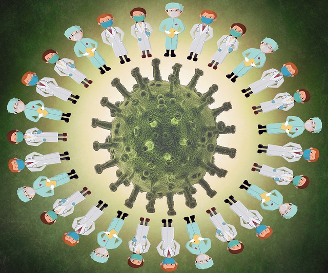
What is Severe Acute Respiratory Syndrome (SARS)?

Severe Acute Respiratory Syndrome (SARS) is a viral respiratory illness caused by the SARS coronavirus (SARS-CoV). It emerged as a global health threat in 2002–2003 when an outbreak originating in China spread to several countries, resulting in thousands of cases and hundreds of deaths.

Causes

  • Severe Acute Respiratory Syndrome (SARS) is caused by a virus known as the SARS coronavirus (SARS-CoV). This virus is a member of the coronavirus family, which includes several other viruses known to cause respiratory illnesses in humans and animals.
  • The SARS-CoV virus is believed to have originated in bats and was transmitted to humans through intermediate animal hosts, such as civet cats, in the early 2000s. The exact mechanism of transmission from animals to humans is not fully understood, but it likely involved close contact with infected animals or their bodily fluids in live animal markets or other settings.
  • Once the virus infects a human host, it can spread from person to person through respiratory droplets produced when an infected individual coughs, sneezes, or talks. Close contact with an infected person, particularly in crowded or poorly ventilated environments, increases the risk of transmission.

The incubation period

The incubation period for severe acute respiratory syndrome (SARS) typically spans from 2 to 10 days. During this phase, individuals may have contracted the SARS coronavirus (SARS-CoV) but do not yet display symptoms of the illness. This period is crucial, as it marks the time between exposure to the virus and the onset of symptoms.

Understanding the duration of the incubation period aids healthcare professionals in identifying and monitoring individuals who may have been exposed to SARS-CoV, facilitating prompt testing, isolation, and preventive measures.

In addition, knowledge of the incubation period assists public health authorities in implementing effective strategies to control the spread of SARS during outbreaks.

The mode of transmission

The mode of transmission for Severe Acute Respiratory Syndrome (SARS) primarily involves droplet infection, facilitated especially by close contact in confined or poorly ventilated environments. Here’s an expanded explanation:

  • Droplet Transmission: SARS-CoV, the virus responsible for SARS, is primarily transmitted through respiratory droplets expelled when an infected individual coughs, sneezes, or talks. These droplets contain viral particles and can travel through the air and infect others who are in close proximity, typically within a distance of about six feet.
  • Air Travelers as Vectors: Air travel plays a significant role in the global spread of SARS. Infected individuals traveling by air can transmit the virus to others on the same flight, particularly if they are in close proximity for an extended period of time. The recirculation of air within aircraft cabins can also contribute to the spread of the virus among passengers.
  • Promotion in Closed Environments: The spread of SARS-CoV is particularly promoted in closed environments where people gather in close quarters, such as airplanes, buses, trains, hospitals, and crowded public spaces. In these settings, respiratory droplets can easily spread from person to person, increasing the risk of transmission.
  • Poor Ventilation: Inadequate ventilation in indoor spaces can exacerbate the spread of SARS-CoV by allowing respiratory droplets to linger in the air for longer periods of time. Proper ventilation, which involves the exchange of indoor air with outdoor air, helps dilute and remove infectious droplets, reducing the risk of transmission.
  • Surface Contamination: While droplet transmission is the primary mode of SARS-CoV transmission, the virus can also spread through contact with contaminated surfaces or objects. Infected individuals may deposit viral particles onto surfaces through respiratory secretions, and others can become infected by touching these surfaces and then touching their mouth, nose, or eyes.
  • Preventive Measures: To mitigate the transmission of SARS-CoV, especially in high-risk environments like healthcare settings and public transportation, preventive measures such as wearing masks, practicing good hand hygiene, maintaining physical distance, and improving ventilation are crucial. These measures help reduce the likelihood of respiratory droplets reaching susceptible individuals and minimize the risk of surface contamination.

Signs and symptoms

The signs and symptoms of severe acute respiratory syndrome (SARS) encompass a range of manifestations that typically emerge following exposure to the SARS coronavirus (SARS-CoV). Here’s an expanded explanation of these symptoms:

  • Fever: A patient with SARS will present with a high fever, typically exceeding 38 degrees Celsius (100.4 degrees Fahrenheit). And also, fever is one of the hallmark symptoms of the illness and may persist for several days.
  • Sneezing: Some patients infected with SARS-CoV may experience sneezing, which is characterized by the sudden, forceful expulsion of air through the nose and mouth. Sneezing can contribute to the spread of respiratory droplets containing the virus to others nearby.
  • Dry Cough: A dry, non-productive cough is a common symptom of SARS patients. This type of cough is characterized by the absence of mucus or phlegm and may be persistent and irritating.
  • Loss of Appetite: SARS can lead to a decreased desire to eat, resulting in a loss of appetite. This symptom may be accompanied by nausea or gastrointestinal discomfort.
  • Weight Loss: Significant weight loss may occur in patients with SARS, particularly due to the combined effects of decreased appetite, fever, and metabolic changes associated with the illness.
  • General Body Malaise: SARS is often associated with a general feeling of illness or malaise, characterized by fatigue, weakness, and overall discomfort. This symptom can significantly impact the individual’s daily activities and quality of life.
  • Headache: Headache is a common complaint among patients with SARS and may vary in intensity from mild to severe. Headaches may be accompanied by other symptoms, such as fever and muscle aches.
  • Joint Pains: Some patients with SARS may experience joint pains, also known as arthralgia. These pains can affect multiple joints throughout the body and may contribute to overall discomfort and mobility issues.

Medical Management

When evaluating a patient suspected of having severe acute respiratory syndrome (SARS), healthcare providers should conduct a comprehensive history and physical examination to assess for relevant symptoms and risk factors. Here’s what can be included.

History

  • Symptoms Onset: Ask the patient about the onset and duration of symptoms, including fever, cough, shortness of breath, and any associated symptoms such as headaches or muscle aches.
  • Travel History: Ask about the patient’s recent travel history, particularly to regions with known SARS outbreaks or areas experiencing community transmission of respiratory illnesses.
  • Exposure History: Determine if the patient has had close contact with people diagnosed with SARS or if they have been in settings where SARS transmission is likely, such as healthcare facilities or crowded public spaces.
  • Occupational History: Assess the patient’s occupation and any potential occupational exposures that may increase the risk of SARS transmission, such as working in healthcare or travel-related industries.
  • Medical History: Review the patient’s medical history, including any underlying health conditions or immunocompromising conditions that may predispose them to severe illness from SARS, such as HIV/AIDS.
  • Contact with Animals: Ask the patient about any recent contact with animals, particularly exotic animals, as certain zoonotic coronaviruses have been associated with animal-to-human transmission.

Physical Examination

  • Vital Signs: Measure the patient’s vital signs, including temperature, pulse rate, respiratory rate, and blood pressure. Elevated temperatures above 37.5 degrees and respiratory rates may indicate fever and respiratory distress, respectively.
  • Respiratory Examination: Perform a thorough respiratory examination, including auscultation of lung sounds, assessment of respiratory effort, and observation for signs of respiratory distress such as tachypnea, dyspnea, or use of accessory muscles.
  • General Appearance: Evaluate the patient’s general appearance and level of consciousness for signs of distress or lethargy. Pallor, diaphoresis, and altered mental status may indicate a severe illness.
  • Examination of Other Systems: Conduct a focused examination of other organ systems, as warranted by the patient’s symptoms and clinical presentation. Pay particular attention to findings suggestive of systemic involvement or complications, such as rash, lymphadenopathy, or signs of multiorgan dysfunction.
  • Skin Examination: Assess the patient’s skin for signs of rash or petechiae, which may indicate an underlying viral infection or coagulopathy.
  • Neurological Examination: Perform a brief neurological examination to evaluate cognitive function, cranial nerve function, motor strength, sensation, and coordination.

Investigations

When evaluating a patient suspected of having Severe Acute Respiratory Syndrome (SARS), several investigations may be conducted to confirm the diagnosis, assess disease severity, and guide management. These investigations typically include the following:

Laboratory Tests

  • Polymerase Chain Reaction (PCR): Nasopharyngeal or throat swab samples are collected and tested using PCR to detect the presence of SARS coronavirus (SARS-CoV) genetic material. This test is crucial for confirming the diagnosis of SARS.
  • Complete Blood Count (CBC): A CBC will reveal leukopenia (low white blood cell count), which is commonly observed in patients with SARS. Thrombocytopenia (low platelet count) and lymphopenia (low lymphocyte count) may also be present.
  • Biochemical Profile: Blood tests may be performed to assess liver and kidney function, electrolyte levels, and markers of inflammation such as C-reactive protein (CRP) and procalcitonin.

Imaging Studies:

  • Chest X-ray: A chest X-ray may be obtained to evaluate for the presence of pneumonia or other pulmonary abnormalities associated with SARS. Findings may include patchy or diffuse infiltrates consistent with viral pneumonia.
  • Computed Tomography (CT) Scan: CT imaging of the chest may provide a more detailed assessment of lung involvement and help differentiate between viral pneumonia and other respiratory conditions.

Viral Culture

  • Viral culture may be attempted to isolate and grow SARS-CoV from respiratory specimens. However, this method is less commonly used due to its lower sensitivity and longer turnaround time compared to PCR testing.

Serological Tests

  • Serological tests measure the presence of antibodies against SARS-CoV in the blood. However, these tests are not typically used for acute diagnosis of SARS due to delayed antibody production during the course of infection.

Other Investigations:

  • Arterial Blood Gas (ABG) Analysis: ABG analysis may be performed to assess respiratory status and oxygenation in patients with severe respiratory distress.
  • Coagulation Studies: Coagulation studies, including prothrombin time (PT) and activated partial thromboplastin time (aPTT), may be indicated to evaluate for coagulopathy associated with severe SARS.

Infection Control Measures:

  • In addition to diagnostic investigations, healthcare providers should implement appropriate infection control measures, including isolation of suspected cases, use of personal protective equipment (PPE), and adherence to strict hand hygiene protocols, to prevent the spread of SARS-CoV in healthcare settings.

Treatment

Treatment for Severe Acute Respiratory Syndrome (SARS) primarily focuses on symptomatic management, supportive care, and infection control measures. Here’s an expanded explanation of treatment options:

Symptomatic Relief

  • Analgesics: Pain relievers such as acetaminophen (e.g., Panadol) can be prescribed to alleviate fever, headache, muscle aches, and other symptoms of discomfort associated with SARS.
  • Nasal Drops: Nasal decongestants or saline nasal drops should be recommended to relieve nasal congestion and facilitate easier breathing, especially in patients experiencing respiratory symptoms.

Respiratory Support

  • Oxygen Therapy: Patients with severe respiratory distress or hypoxemia may require supplemental oxygen therapy to maintain adequate oxygenation levels and alleviate breathing difficulties.
  • Mechanical Ventilation: In severe cases of SARS with respiratory failure, invasive mechanical ventilation via endotracheal intubation may be necessary to support breathing and ensure adequate gas exchange.
  • Tracheostomy: In rare instances where prolonged mechanical ventilation is required or if there are complications related to endotracheal intubation, a tracheostomy procedure may be performed to establish a secure airway.

Antimicrobial Therapy

  • Broad-Spectrum Antibiotics: While SARS is caused by a virus, bacterial coinfections or secondary bacterial pneumonia can occur, particularly in hospitalized patients. Broad-spectrum antibiotics may be prescribed empirically to prevent or treat bacterial infections and reduce the risk of complications.

Antiviral Therapy

  • Antiviral Medications: Although no specific antiviral drugs have been approved for the treatment of SARS-CoV infection, experimental antiviral agents such as ribavirin, lopinavir/ritonavir, and interferons have been investigated in clinical trials for their potential efficacy in managing SARS.

Isolation and Infection Control

  • Isolation Precautions: Patients diagnosed with SARS should be placed in airborne isolation rooms or designated isolation areas to prevent the transmission of the virus to healthcare workers and other patients.
  • Personal Protective Equipment (PPE): Healthcare providers caring for patients with SARS should adhere to strict infection control protocols, including the use of appropriate PPE such as N95 respirators, gloves, gowns, and eye protection.

Supportive Care

  • Fluid and Nutritional Support: Adequate hydration and nutritional support are essential for patients with SARS to maintain hydration, support immune function, and promote recovery.
  • Monitoring and Surveillance: Close monitoring of vital signs, respiratory status, laboratory parameters, and clinical progression is essential to detect any deterioration in the patient’s condition and adjust treatment accordingly.

Experimental Therapies

  • Clinical Trials: Patients with severe or refractory SARS may be eligible to participate in clinical trials evaluating novel therapeutic agents or investigational treatments for SARS-CoV infection.

Nursing Management

Patient Isolation

  • Implement strict airborne isolation precautions for patients diagnosed with SARS.
  • Ensure that patients are placed in negative-pressure rooms with appropriate ventilation.
  • Provide education to patients and visitors about the importance of adhering to isolation protocols, and make sure the room where the patient with SARS is marked.
  • Use personal protective equipment (PPE), such as N95 respirators, gloves, gowns, and eye protection, when entering the patient’s room.

Infection Prevention

  • A nurse should promote hand hygiene practices among healthcare staff, patients, and visitors.
  • A nurse should implement environmental cleaning and disinfection protocols for surfaces and equipment.
  • A nurse should monitor and enforce compliance with infection control measures.
  • A nurse should educate patients and families about respiratory etiquette, including covering coughs and sneezes with tissues or elbows.

Disease Notification

  • A nurse should notify public health authorities promptly of suspected or confirmed cases of SARS.
  • A nurse should provide accurate and timely information to healthcare staff, patients, and families about the status of SARS cases and any updates to infection control measures.
  • A nurse should collaborate with local health departments to facilitate contact tracing and outbreak investigation.
  • A nurse should ensure the confidentiality of patient information in accordance with healthcare privacy regulations.

Maintenance of the Airway

  • A nurse should assess and monitor the patient’s respiratory status, including respiratory rate, effort, and oxygen saturation, using a pulse oximeter.
  • Make sure to position the patient at a 90-degree angle to comfortably optimize airway patency and lung expansion.
  • Administer supplemental oxygen therapy as prescribed to maintain adequate oxygenation, if oxygen levels are below 95%.
  • Initiate respiratory treatments such as nebulization or chest physiotherapy to assist with airway clearance, if indicated.

Maintenance of Breathing

  • A nurse should monitor respiratory rate, depth, and pattern regularly to prevent respiratory failure.
  • A nurse should encourage deep breathing exercises and coughing to mobilize secretions and maintain lung function.
  • Administer prescribed bronchodilators or mucolytic agents to improve airway clearance and breathing comfort.
  • Provide respiratory support, such as non-invasive ventilation, if respiratory distress worsens.

Maintenance of Circulation

  • Monitor vital signs closely, including blood pressure, heart rate, and peripheral perfusion.
  • Assess fluid balance and intervene as needed to maintain hydration and electrolyte balance.
  • Administer intravenous fluids and medications as prescribed to support circulation and manage hemodynamic stability.
  • Collaborate with the healthcare team to address any signs of cardiovascular compromise promptly.

Environment

  • Maintain a clean and well-ventilated environment to reduce the risk of transmission.
  • Ensure adequate spacing between patient beds to minimize close contact and promote social distancing.
  • Make sure the ward is free from noise to reduce noise and promote restful sleep for patients.
  • Monitor and control environmental factors such as temperature and humidity to optimize patient comfort and recovery.

Observation

  • Conduct frequent assessments of the patient’s clinical status, including respiratory status, vital signs, and neurological status.
  • Monitor for any signs of respiratory distress, deterioration, or complications, such as pneumonia or acute respiratory distress syndrome (ARDS).
  • Document and communicate findings accurately to the healthcare team to facilitate appropriate interventions and decision-making.
  • Use standardized assessment tools and scoring systems as appropriate to track changes in patient conditions over time.

Psychological Care

  • Provide emotional support and reassurance to patients and families experiencing anxiety or distress.
  • Encourage open communication and active listening to address concerns and fears related to the illness.
  • Offer psychological interventions such as relaxation techniques, mindfulness exercises, or counseling referrals as needed.
  • Collaborate with interdisciplinary team members, including social workers or psychologists, to address psychosocial needs effectively.

Hygiene

  • Promote adherence to hand hygiene practices among healthcare staff, patients, and visitors.
  • Provide education on proper handwashing technique and the use of hand sanitizer.
  • Ensure the availability of hand hygiene supplies at all points of care.
  • Monitor and reinforce compliance with hygiene protocols through regular audits and feedback.

Nutrition

  • Assess the patient’s nutritional status and dietary intake regularly.
  • Provide nutritional counseling and support to ensure adequate calorie and protein intake to promote quick healing.
  • Offer small, frequent meals and snacks to maintain energy levels and prevent malnutrition.
  • Collaborate with dietitians or nutritionists to develop individualized meal plans and address any dietary restrictions or preferences.

Elimination

  • Monitor urinary output and bowel function regularly.
  • Encourage adequate fluid intake to maintain hydration and promote urinary output.
  • Assist patients with toileting needs and provide appropriate hygiene care.
  • Implement measures to prevent urinary retention or constipation, such as regular toileting schedules and mobility promotion.

Medication

  • Administer prescribed medications accurately and timely, ensuring adherence to dosing schedules.
  • Monitor for potential drug interactions or adverse effects of medications.
  • Educate patients about the purpose, dosage, and side effects of prescribed medications.
  • Document medication administration and response to treatment in the patient’s medical record.

Advice at Discharge

  • Provide education and support to patients and families facing social or economic disadvantages.
  • Assist with access to community resources such as social services, financial assistance programs, or support groups.
  • Advocate for patients’ needs and rights within the healthcare system.
  • Collaborate with case managers or social workers to address barriers to care and promote health equity.

Read more

Coryza | Causes | Pathophysiology | Signs and symptoms | Treatment | Nursing Management

What is coryza?

“Coryza” is a medical term referring to inflammation of the mucous membranes in the nose, typically associated with symptoms such as congestion, runny nose, sneezing, and sometimes mild fever. It’s a common symptom of the common cold or other respiratory infections.

Cause

  • Viral infections: The common cold is the most frequent cause of coryza. Other viruses, such as influenza (flu) and respiratory syncytial virus (RSV), can also lead to coryza symptoms.
  • Bacterial infections: While less common than viral infections, bacterial infections such as sinusitis or bacterial pneumonia can also cause coryza.
  • Allergies: Allergic rhinitis, caused by allergens such as pollen, dust mites, or pet dander, can result in coryza symptoms.
  • Environmental irritants: Exposure to irritants like smoke, pollution, or strong odors can irritate the nasal passages and lead to coryza.
  • Other factors: Coryza can also be a symptom of certain medical conditions, such as vasomotor rhinitis or hormonal changes during pregnancy.

Mode of transmission

Coryza can spread through various modes of transmission. It is commonly transmitted through direct contact with an infected person, such as shaking hands or touching surfaces contaminated with nasal or throat secretions.

Additionally, the virus or bacteria responsible for coryza can be transmitted through the inhalation of airborne droplets expelled when an infected person coughs, sneezes, or talks.

Indirect transmission is also possible, where the virus or bacteria can survive on surfaces for a period of time. Individuals can contract coryza by touching contaminated surfaces, such as doorknobs, countertops, or shared items like utensils or electronic devices, and then touching their nose or mouth.

Furthermore, coryza can be transmitted through fomites, objects, or articles that have been freshly soiled by the nasal or throat secretions of infected individuals. This includes items like tissues, handkerchiefs, towels, or shared personal items.

Pathophysiology

The pathophysiology of coryza involves a series of events that lead to the characteristic symptoms of nasal congestion, runny nose, and throat discomfort. It typically begins with the invasion of a virus or bacteria into the upper respiratory tract.

Once inside the body, these pathogens trigger an immune response, leading to inflammation of the mucous membranes lining the nose, pharynx, and tonsils. This inflammation causes the blood vessels in the affected tissues to dilate, resulting in swelling and congestion.

Simultaneously, the immune system releases various chemical mediators, such as histamine, which further exacerbate the inflammation and increase mucus production. The excess mucus accumulates in the nasal passages, leading to a runny nose and postnasal drip.

Further, the swelling and inflammation can block the openings of the sinuses, impairing their drainage and leading to sinus congestion and pressure. This can contribute to symptoms such as facial pain or headaches.

Overall, the pathophysiology of coryza involves a complex interplay between viral or bacterial invasion, immune response, inflammation, and mucus production, ultimately resulting in the hallmark symptoms of nasal congestion, runny nose, and throat irritation.

Signs and symptoms

The signs and symptoms of coryza encompass a range of discomforts typically experienced during the course of the illness:

  1. Runny nose and excessive tearing (lacrimation): A patient with coryza often experiences a profuse discharge of fluid from the nose, accompanied by increased tear production, leading to watery eyes.
  2. Nasal discharge: Alongside excessive tearing, there is typically a discharge from the nose, which can vary in consistency from thin and watery to thicker and mucoid.
  3. General body malaise: Coryza is often accompanied by a general feeling of unwellness, characterized by fatigue, weakness, and a sense of discomfort throughout the body. This malaise can persist for a duration of 2–7 days, depending on the severity of the infection.
  4. Nasal congestion: swelling and inflammation of the nasal passages lead to congestion, causing difficulty breathing through the nose.
  5. Sneezing: The irritation and inflammation of the nasal mucosa can trigger frequent bouts of sneezing, which serve as a reflexive response to expel irritants from the nasal passages.
  6. Sore throat: Inflammation of the throat mucosa commonly results in throat discomfort, pain, and difficulty swallowing, contributing to a sore throat.
  7. Body chills: Individuals with coryza may experience episodes of chills or shivering, often accompanied by a mild fever, as the body attempts to fight off the infection.
  8. Headache: Sinus congestion, inflammation, and pressure can lead to headaches, which are commonly experienced as a dull ache or pressure in the forehead, temples, or behind the eyes.
  9. Coughing: As irritation and inflammation extend to the lower respiratory tract, individuals may develop a cough, which can be dry or productive, as the body attempts to clear mucus and irritants from the airways.

Medical Management

In taking the history of a patient presenting with symptoms suggestive of coryza, the healthcare provider will typically ask about:

History Taking

  • Onset of Symptoms: When did the symptoms start? Are they sudden or gradual in onset?
  • Duration of Symptoms: How long have the symptoms been present?
  • Symptom Description: What specific symptoms are experienced? (Runny nose, nasal congestion, sneezing, sore throat, coughing, fever, headache, etc.)
  • Severity of Symptoms: How severe are the symptoms? Are they interfering with daily activities?
  • Associated Symptoms: Are there any other symptoms present, such as body aches, fatigue, or loss of appetite?
  • Exposure History: Have there been any recent exposures to sick individuals, crowded places, or travel to areas with known outbreaks?
  • Medical History: Does the patient have any underlying medical conditions or allergies? Are they taking any medications?
  • Social History: What is the patient’s occupation and living environment? Do they smoke or have regular exposure to environmental pollutants?
  • Allergy History: Does the patient have a history of allergies to pollen, dust, pets, or other allergens?

Physical Examination

  • Vital Signs: Measure temperature, heart rate, respiratory rate, and blood pressure.
  • Head and Neck Examination:
    • Inspect the nose for signs of nasal congestion, discharge, or inflammation.
    • Evaluate the throat for redness, swelling, or exudate on the tonsils.
    • Palpate the neck lymph nodes for enlargement or tenderness.
  • Respiratory Examination:
    • Auscultate lung sounds to assess for signs of congestion, wheezing, or abnormal breath sounds.
    • Evaluate for signs of respiratory distress, such as increased work of breathing or use of accessory muscles.
  • Eye Examination: Look for signs of conjunctivitis (redness, discharge) if present.
  • Skin Examination: Check for rashes or other skin abnormalities if indicated.
  • Additional Examinations: Perform any additional examinations based on the patient’s symptoms and clinical presentation.

Investigations

In the diagnosis of coryza, investigations may be conducted to confirm the underlying cause and rule out other potential conditions. Common investigations include:

  • Nasal Swab: A sample of nasal secretions should be collected using a swab to test for the presence of viral or bacterial pathogens, particularly if a nurse provider suspects a specific infectious cause such as influenza or bacterial sinusitis.
  • Complete Blood Count (CBC): A blood test may be performed to assess for signs of infection, such as an elevated white blood cell count, which can indicate an active immune response to infection.
  • Throat Swab: In cases where a sore throat is a prominent symptom, a swab of the throat may be collected to test for the presence of bacteria, such as group A Streptococcus, which can cause strep throat.
  • Chest X-ray: If the patient presents with symptoms suggestive of lower respiratory tract involvement, such as persistent cough, chest pain, or difficulty breathing, a chest X-ray may be ordered to evaluate for signs of pneumonia or other pulmonary conditions.
  • Allergy Testing: For patients with recurrent or chronic coryza symptoms, allergy testing may be performed to identify specific allergens triggering the allergic response. This can include skin prick tests or blood tests (such as IgE antibody testing).
  • Nasal Endoscopy: In cases of chronic or severe nasal congestion or sinusitis, a nasal endoscopy may be performed to visualize the nasal passages and sinuses and assess for structural abnormalities or signs of chronic inflammation.
  • Viral Testing: In outbreaks or cases where a specific viral cause is suspected, such as during influenza season, molecular tests such as polymerase chain reaction (PCR) may be used to detect viral RNA or DNA in respiratory samples.
  • Culture and Sensitivity Testing: In cases where bacterial infection is suspected, cultures of nasal or throat swabs may be performed to identify the specific bacteria involved and determine their sensitivity to antibiotics.

Treatment

The treatment of coryza usually involves managing symptoms and supporting the body’s immune response. While there is no specific cure for coryza, the following measures can help alleviate discomfort and promote recovery:

  • Hydration: A nurse should encourage the patient to drink plenty of fluids, such as water, herbal teas, and clear soups. Adequate hydration helps thin mucus secretions, making it easier to clear congestion and maintain hydration levels.
  • Salt Gargles: Telling the patient to take some salt gargles can help soothe a sore throat and reduce inflammation. Mix one teaspoon of salt in one liter of warm water and instruct the patient to gargle with the solution several times a day.
  • Anti-inflammatory Drugs: Nonsteroidal anti-inflammatory drugs (NSAIDs) like aspirin can help reduce inflammation and alleviate pain. A typical regimen may include aspirin, 600 mg, taken three times a day for three days. However, caution should be exercised with aspirin use, particularly in children and individuals with certain medical conditions, due to the risk of Reye’s syndrome and other adverse effects.
  • Antihistamines: Antihistamines such as Piriton (chlorphenamine) can help relieve symptoms like sneezing, runny nose, and itching by blocking the action of histamine. The recommended dosage is Piriton 4 mg, taken twice a day for three days. Antihistamines can cause drowsiness, so patients should avoid activities requiring alertness until they know how the medication affects them.
  • Decongestants: Decongestants help reduce nasal congestion by constricting blood vessels in the nasal passages, thus drying up secretions and relieving stuffiness. Over-the-counter nasal decongestants or oral formulations may be used as directed.
  • Nose Drops: Nasal drops containing ephedrine (for adults) or normal saline (for children) can help relieve nasal congestion and promote the drainage of mucus. These drops should be administered according to the instructions provided and should not be used for prolonged periods to avoid rebound congestion.
  • Increased Vitamin C Intake: Vitamin C is thought to support the immune system and may help reduce the duration and severity of respiratory infections. Encourage the patient to consume vitamin C-rich foods such as citrus fruits, berries, and leafy greens, or consider supplementation if appropriate.
  • Bed Rest: Adequate rest is essential for supporting the body’s immune response and promoting recovery from coryza. Encourage the patient to get plenty of rest and avoid strenuous activities until symptoms improve.

Nursing Management for Coryza

Aims

  1. Symptom relief
  2. Prevention of complications
  3. Promotion of recovery
  4. Patient education and empowerment

Patient Isolation

  • Place the patient in a private room with the door closed to prevent the spread of respiratory droplets. Ensure that healthcare personnel wear appropriate personal protective equipment (PPE), including masks, gloves, and gowns, when in close contact with the patient.
  • Inform the patient and his or her family visitors about the importance of hand hygiene and respiratory etiquette, including covering the mouth and nose when coughing or sneezing, to reduce the transmission of infectious respiratory droplets.
  • Make sure you restrict non-essential visitors and encourage virtual communication methods to minimize contact and prevent the spread of the virus to others.
  • The nurses should ensure thorough cleaning and disinfection of patient care areas and equipment to prevent contamination and transmission of the virus to other patients and healthcare workers.

Infection Prevention

  • Make an emphasis on the importance of frequent handwashing with soap and water or alcohol-based hand sanitizers for both the patient and healthcare personnel to prevent the spread of infection.
  • Educate the patient on proper respiratory etiquette, including covering the mouth and nose when coughing or sneezing with a tissue or the elbow, and disposing of used tissues promptly in a lined waste bin.
  • Maintain adequate ventilation in patient rooms and ensure proper disposal of contaminated materials to minimize the risk of airborne transmission by opening up nearby windows.
  • Monitor adherence to isolation precautions among healthcare personnel and provide ongoing education and reinforcement to ensure compliance with infection control protocols.

Maintenance of the Airway

  • Make sure to position the patient to sit upright or elevate the head of the bed at a 90-degree angle to facilitate drainage of nasal secretions and improve breathing comfort.
  • Use humidifiers or steam inhalation to moisten the air and help loosen thick nasal secretions, making it easier for the patient to clear their airway.
  • Administer saline nasal drops to lubricate and hydrate nasal passages, facilitating the removal of mucus and relieving nasal congestion.
  • Instruct the patient to perform deep breathing exercises to maintain lung expansion and prevent respiratory complications such as atelectasis or pneumonia.

Maintenance of Breathing

  • A nurse should regularly assess the patient’s respiratory rate and pattern to detect any signs of respiratory distress or deterioration.
  • Administer supplemental oxygen if the oxygen levels are below 95% as prescribed to maintain adequate oxygenation and alleviate respiratory distress if hypoxemia is present.
  • Teach effective coughing techniques, such as huffing or controlled coughing, to help clear mucus from the airways and improve breathing.
  • If necessary, perform nasopharyngeal or oropharyngeal suctioning to remove excess mucus and maintain a clear airway, especially in patients with difficulty expecting secretions.

Maintenance of Circulation

  • A nurse should regularly monitor the patient’s vital signs, including heart rate, blood pressure, and temperature, to detect any signs of hemodynamic instability or complications such as dehydration.
  • Encourage the patient to take adequate fluid intake to prevent dehydration, especially if the patient has a fever or is experiencing increased respiratory secretions.
  • Assess peripheral perfusion by monitoring skin color, temperature, and capillary refill time to evaluate circulatory status and detect signs of inadequate tissue perfusion.
  • Encourage the patient to do some early ambulation and frequent position changes to promote venous return and prevent complications such as venous thromboembolism (VTE) associated with immobility.

Environment

  • Make sure that the ward is a clean and clutter-free environment to reduce the risk of infection transmission and promote patient comfort and well-being.
  • Make sure the patient is comfortable at room temperature by switching on heaters to promote patient comfort and prevent excessive cooling or overheating, which can exacerbate respiratory symptoms.
  • Make sure the ward is free from noise and distractions in the patient’s environment to facilitate rest and promote relaxation, which is essential for recovery.
  • Encourage personalization of the patient’s environment with familiar belongings or comforting items to provide a sense of security and enhance emotional well-being.

Observation

  • Conduct frequent assessments of the patient’s vital signs, respiratory status, and overall condition to detect any changes or deterioration promptly.
  • Document all observations, interventions, and patient responses accurately and promptly in the medical record to facilitate communication and continuity of care.
  • Evaluate the patient’s response to nursing interventions and treatments to determine their effectiveness and make necessary adjustments to the plan of care.
  • Communicate relevant findings and concerns to the healthcare team, including physicians, respiratory therapists, and other interdisciplinary team members, to ensure coordinated and comprehensive care.

Psychological Care

  • Provide emotional support and reassurance to the patient and their family members, acknowledging their concerns and addressing any fears or anxieties related to the illness.
  • Listen attentively to the patient’s feelings and concerns, validating their experiences and providing empathy and understanding.
  • Encourage the patient to express their emotions and discuss their coping strategies, offering encouragement and positive reinforcement.
  • Identify patients at risk of psychological distress or crisis and provide appropriate interventions, including referral to mental health professionals if needed.

Hygiene

  • Assist the patient with personal hygiene activities, such as bathing, oral care, and changing clothes, to maintain cleanliness and promote comfort.
  • Ensure cleanliness and hygiene in the patient’s environment, including regular cleaning and disinfection of surfaces, linens, and equipment.
  • Reinforce the importance of hand hygiene for the patient and their caregivers, providing education on proper handwashing techniques and the use of hand sanitizers.
  • Adhere to standard precautions and infection control protocols, including the use of personal protective equipment (PPE), to prevent the spread of infection within the healthcare setting.

Nutrition

  • Assess the patient’s nutritional status and dietary intake, identifying any deficits or nutritional needs that may impact their recovery.
  • Encourage the patient to eat a balanced diet rich in nutrients, including fruits, vegetables, lean proteins, and whole grains, to support immune function and promote healing.
  • If necessary, collaborate with the healthcare team to provide nutritional support, such as oral nutritional supplements or enteral feeding, to ensure adequate caloric and nutrient intake.
  • Monitor the patient’s fluid intake and output closely, ensuring adequate hydration to prevent dehydration and support respiratory function.

Elimination

  • Assist the patient with toileting needs, ensuring accessibility and privacy, especially for patients with mobility limitations or urinary incontinence.
  • Monitor urinary output and assess for signs of urinary retention or dehydration, intervening as needed to promote adequate hydration and urinary function.
  • Encourage the patient to maintain regular bowel habits through dietary measures, adequate fluid intake, and physical activity to prevent constipation and discomfort.
  • Provide appropriate management strategies for patients with urinary or fecal incontinence, including the use of absorbent pads or briefs and implementing toileting schedules.

Medication

  • Administer prescribed medications promptly and accurately, ensuring adherence to the prescribed dosage, route, and frequency.
  • Provide thorough medication education to the patient and their caregivers, including information on medication purpose, dosage regimen, potential side effects, and precautions.
  • Monitor the patient for any adverse reactions or side effects related to medication administration, reporting any concerns to the healthcare provider promptly.
  • Conduct medication reconciliation regularly to ensure accurate and up-to-date medication lists, minimizing the risk of medication errors and drug interactions.

Advice at Discharge

  • Follow-up Care: Provide instructions for follow-up care, including any necessary appointments with healthcare providers or specialists for ongoing management of the patient’s condition.
  • Home Care Instructions: Educate the patient and their caregivers on home care strategies, including symptom management, medication administration, and when to seek medical attention if symptoms worsen or new symptoms develop.
  • Preventive Measures: Reinforce the importance of preventive measures, such as hand hygiene, respiratory etiquette, and vaccination, to reduce the risk of recurrent respiratory infections.
  • Community Resources: Provide information on community resources and support services available to the patient and their family, including support groups, home health services, and other healthcare resources for continued assistance and support post-discharge.

Complications

Complications associated with coryza may involve various parts of the respiratory and adjacent systems:

  • Lower Respiratory Tract Infections: Coryza can progress to affect the lower respiratory tract, leading to conditions such as bronchitis or pneumonia. This progression may occur when the infection spreads from the upper respiratory tract to the bronchi and lungs, causing inflammation and potentially severe respiratory symptoms.
  • Sinusitis: Inflammation of the sinuses, known as sinusitis, can occur as a complication of coryza. The inflammation may result from blockage of the sinus openings due to swollen nasal passages and excess mucus production, leading to sinus pressure, facial pain, and nasal congestion.
  • Otitis Media: Particularly common in children, coryza can lead to otitis media, an infection of the middle ear. The blockage of the Eustachian tube, which connects the middle ear to the throat, can promote the accumulation of fluid and bacteria in the middle ear, resulting in pain, earache, and potential complications such as abscess formation.
  • Tonsillitis: Infection of the tonsils, known as tonsillitis, can occur concurrently with or as a complication of coryza. The inflamed tonsils may become swollen, red, and painful, leading to difficulty swallowing, a sore throat, and sometimes fever.
  • Bronchitis: Coryza can lead to inflammation of the bronchial tubes, resulting in bronchitis. Symptoms may include coughing, chest discomfort, and difficulty breathing. Bronchitis can be acute or chronic, depending on the duration and severity of symptoms.
  • Pneumonia: More common in vulnerable populations such as infants and the elderly, coryza can predispose individuals to pneumonia. Infection of the lungs can lead to symptoms such as fever, cough, chest pain, and difficulty breathing. Pneumonia may be caused by viral or bacterial pathogens, and severe cases may require hospitalization and intensive treatment.

Read more