Cardiac arrest can happen anywhere on a hospital unit, in a clinic hallway, at a sporting event, or in a living room. Survival hinges on immediate action that maintains circulation and oxygen delivery until defibrillation and advanced care arrive. That action is CPR. But what does CPR stand for, and why does it work? This comprehensive guide, written in a clinical, educational tone, defines cardiopulmonary resuscitation, breaks down the science behind it, clarifies step-by-step priorities, and reviews quality metrics, special situations, training pathways, and common myths.
What Does CPR Stand For | Cardiopulmonary Resuscitation Meaning, Steps, Quality, Certification
Note: Educational content only. Follow local protocols, prescriber orders, and certifying-body guidelines. In emergencies, activate the local emergency response system without delay.
Quick Answer-What CPR Stands For
- CPR = Cardiopulmonary Resuscitation.
- Cardio: the heart’s pumping function.
- Pulmonary: the lungs’ oxygenation and ventilation.
- Resuscitation: restoring perfusion and breathing to sustain life.
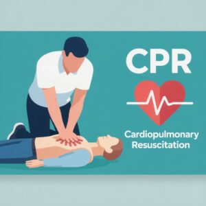
Cardiopulmonary resuscitation is a coordinated set of interventions chiefly chest compressions, ventilations, and early defibrillation that maintains blood flow and oxygenation during cardiac arrest or severe respiratory failure until return of spontaneous circulation (ROSC) or advanced interventions occur.
Why CPR Matters: Chain of Survival
Resuscitation science highlights the Chain of Survival. Strong links improve outcomes across in-hospital and out-of-hospital settings:
- Early recognition and emergency activation
- Immediate high-quality CPR with minimal interruptions
- Rapid defibrillation for shockable rhythms
- Advanced life support and postarrest care
- Recovery support and rehabilitation
Each link depends on the previous one; high-quality CPR buys time for defibrillation and definitive treatment.
Breaking Down the Term: Cardio-Pulmonary- Resuscitation
Cardio (Heart)
The heart provides perfusion pressure to deliver oxygen and nutrients. In arrest, compressions substitute for the heart’s pump, generating forward blood flow, raising coronary perfusion pressure, and preserving organ function.
Pulmonary (Lungs)
The lungs facilitate oxygen uptake and carbon dioxide removal. Ventilation in CPR supports oxygen delivery and CO2 clearance; in some early minutes of adult arrest, chest recoil also draws in limited air, which is why compression-only CPR can be effective for untrained rescuers.
Resuscitation (Restore)
Resuscitation seeks to restore circulation and breathing. Chest compressions provide mechanical circulation; defibrillation treats shockable rhythms (ventricular fibrillation and pulseless ventricular tachycardia); ventilations provide oxygenation and CO2 removal; vasoactive medications and advanced airways are added by trained teams.
A Short History of CPR
- 1950s–1960s: External chest compressions and mouth-to-mouth ventilation enter practice; early defibrillation studies demonstrate survival gains.
- 2000s: Guidelines consolidate around CAB (compressions-airway-breathing), compression-first emphasis, and minimized interruptions.
- 2010s–present: Focus intensifies on compression quality metrics, feedback devices, and low-dose/high-frequency training; widespread deployment of AEDs; enhanced postarrest care.
Core Components of CPR
Chest Compressions
- Rate: 100–120 compressions per minute.
- Depth: Adults at least 2 inches (5 cm) and not more than 2.4 inches (6 cm).
- Recoil: Allow full chest recoil after each compression.
- Fraction: Minimize pauses; aim for chest compression fraction >60–80%.
- Rotation: Switch compressors about every 2 minutes or sooner if fatigued.
Why it works: Adequate rate and depth generate perfusion pressure to maintain coronary and cerebral blood flow, improving odds of ROSC.
Ventilations (Rescue Breaths)
- Single rescuer adult: 30 compressions to 2 breaths (30:2).
- Two-rescuer child/infant: 15:2.
- Advanced airway in place: deliver 1 breath every 6 seconds (10/min) without pausing compressions.
- Avoid hyperventilation; deliver just enough to see chest rise.
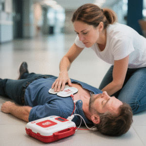
Defibrillation and AED Use
- Defibrillation treats ventricular fibrillation (VF) and pulseless ventricular tachycardia (pVT).
- Apply AED/defibrillator quickly; follow prompts; resume compressions immediately after shock.
- Pad placement: anterolateral (right upper chest, left lateral chest) or anteroposterior as indicated.
Airway and Oxygen
- Basic: head-tilt, chin-lift; jaw thrust if trauma suspected; bag-mask with tight seal and HEPA filter where available.
- Advanced: supraglottic airway or endotracheal tube per trained providers; capnography guides quality (ETCO2 ≥ 10–20 mmHg suggests better perfusion during CPR).
Hands-Only CPR vs Conventional CPR
- Hands-only CPR: chest compressions without rescue breaths; recommended for untrained bystanders in adult sudden cardiac arrest.
- Conventional CPR: compressions plus breaths; preferred for trained rescuers and for pediatric, drowning, asphyxial arrests, or prolonged downtime where ventilation is critical.
Adult vs Pediatric vs Infant Modifications
Adults (Puberty and Older)
- Compression depth: ≥2 inches (5 cm), not >2.4 in (6 cm).
- Hand position: center of chest on lower half of sternum; two hands.
- Ratio: 30:2 for single rescuer; advanced airway enables continuous compressions with asynchronous breaths.
Children (1 Year to Puberty)
- Compression depth: about 2 inches (5 cm) or one-third anterior–posterior chest diameter.
- Hands: one or two hands depending on size.
- Ratio: 30:2 single rescuer; 15:2 with two rescuers.
Infants (Under 1 Year)
- Compression depth: about 1.5 inches (4 cm) or one-third AP chest diameter.
- Technique:
- Single rescuer: two fingers just below nipple line on sternum.
- Two rescuers: two-thumb encircling technique for superior perfusion.
- Ratio: 30:2 single rescuer; 15:2 two rescuers.
- Ventilations: gentle puffs sufficient to see chest rise; avoid overdistension.
Special Situations
Drowning and Asphyxial Arrest
- Emphasize early ventilations; if trained, provide 2 rescue breaths before compressions.
- Remove from water and place on firm surface before starting compressions.
Opioid-Related Emergencies
- If unresponsive and not breathing normally: begin CPR; administer naloxone when available; do not delay compressions.
Trauma
- Prioritize safety, hemorrhage control, and airway with spinal precautions; consider reversible causes (tension pneumothorax, tamponade).
Pregnancy
- Standard hand position; perform manual left uterine displacement to relieve aortocaval compression when fundus is at/above umbilicus.
- Early activation of obstetric and neonatal teams; perimortem delivery protocols per institutional policy when indicated.
Hypothermia
- Check for signs of life with care; begin CPR; avoid aggressive limb movement; defibrillation and medications may be less effective until warmed.
Choking (Foreign-Body Airway Obstruction)
- Adults/children: abdominal thrusts for severe obstruction in responsive victims; back blows and chest thrusts for infants.
- If unresponsive: start CPR, check mouth for visible object before breaths.
CPR Quality and Team Dynamics
High-quality CPR depends on technique and teamwork:
- Assign roles: compressor, airway/ventilation, AED/defibrillator, medication/recording, team leader.
- Use closed-loop communication: clear orders, repeat-backs, time checks every 2 minutes.
- Monitor quality:
- Real-time feedback devices (rate/depth/recoil)
- Capnography: ETCO2 trend during compressions; sudden rise may indicate ROSC
- Pulse checks only when rhythm and perfusion are likely; keep interruptions under 10 seconds.
AED Essentials-Step by Step
- Turn on AED immediately upon arrival.
- Expose chest; dry if wet; remove medication patches and wipe area.
- Apply pads as illustrated; press firmly for adhesion.
- Follow prompts: “Analyzing”-ensure no one touches the patient.
- If advised, deliver shock, then resume compressions immediately for 2 minutes before next analysis.
- Continue until advanced help takes over or signs of life appear.
Evidence Corner-Why These Steps Work
- Coronary perfusion pressure correlates with ROSC; uninterrupted compressions sustain pressure.
- Early defibrillation doubles to triples survival for VF/pVT.
- Ventilation strategy balances oxygenation with prevention of intrathoracic pressure spikes that reduce venous return.
- Team dynamics and feedback devices improve adherence to rate, depth, and fraction targets.
Infection Prevention in CPR
- Scene safety first; use gloves and eye protection when available.
- Barrier devices: pocket mask or bag-mask with HEPA filter reduce aerosol exposure.
- For lay responders with no barrier, compression-only CPR is acceptable for adults until equipment or trained responders arrive.
Training and Certification Pathways
BLS (Basic Life Support) for Healthcare Providers
- Core content: adult/child/infant CPR with AED, bag-mask ventilation, choking relief, team dynamics.
- Validity: typically 2 years (AHA, ARC, HSI/ASHI); many institutions add low-dose/high-frequency refreshers.
ACLS (Advanced Cardiovascular Life Support)
- Adds rhythm recognition, pharmacology, advanced airways, cardioversion/defibrillation strategies, and leadership.
- Validity: typically 2 years.
PALS (Pediatric Advanced Life Support)
- Pediatric assessment, respiratory failure/shock algorithms, pediatric arrest management.
- Validity: typically 2 years.
RQI and Competency Programs
- Quarterly micro-skill practice on feedback manikins sustains quality between renewals; commonly used in hospital systems.
Legal and Ethical Considerations
- Good Samaritan laws: many jurisdictions protect lay rescuers who render aid in good faith; provisions vary by state and role.
- Duty to act: healthcare professionals on duty follow institutional policy and scope.
- DNR/AND orders: honor established directives in clinical settings; know how to identify and verify them; when in doubt in the community, initiating CPR is often appropriate until advanced directives are clarified.
Common Mistakes and How to Correct Them
- Shallow compressions- focus on 2–2.4 inch depth in adults; use body weight; verify with feedback device.
- Slow rate or excessive rate – target 100–120/min; use a metronome.
- Incomplete recoil- consciously release pressure each time; consider hand repositioning or rotation.
- Prolonged pauses-charge defibrillator during compressions; preassign roles; resume compressions immediately post-shock.
- Hyperventilation- deliver just enough to see chest rise; for advanced airway, ventilate at 10/min without pausing compressions.
- Delayed AED use- send a runner early; integrate AED application while compressions continue.
Myths vs Facts
- Myth: CPR restarts the heart.
- Fact: CPR maintains blood flow; defibrillation and advanced interventions treat lethal arrhythmias. CPR buys time for definitive treatment.
- Myth: Rib fractures mean CPR was done incorrectly.
- Fact: Rib fractures can occur even with correct technique, especially in older adults. The priority is life-saving circulation.
- Myth: Only medical professionals should perform CPR.
- Fact: Lay rescuers can and should provide hands-only CPR while activating emergency services and retrieving an AED.
- Myth: AEDs are complicated and dangerous to use.
- Fact: AEDs are designed with voice prompts and safety checks; they shock only when a shockable rhythm is detected.
Related Clinical Learning for Nurses
Clinical education gains depth when integrated across systems. Topics like airway management, postarrest care, and respiratory conditions reinforce resuscitation readiness. For respiratory-focused study planning, a bronchitis nursing diagnosis care plan complements CPR knowledge by sharpening assessment of ventilation, gas exchange, and airway clearance skills that translate to early recognition of deterioration before arrest.
Glossary of CPR Terms
- ROSC: Return of Spontaneous Circulation; restoration of a palpable pulse and effective blood pressure.
- VF/pVT: Ventricular fibrillation/pulseless ventricular tachycardia; shockable rhythms.
- Asystole/PEA: Non-shockable rhythms requiring high-quality CPR and treatment of reversible causes.
- ETCO2: End-tidal carbon dioxide; surrogate marker for perfusion during CPR.
- Chest Compression Fraction (CCF): Proportion of arrest time spent actively compressing the chest.
Practical Checklist for Readiness
- Know the nearest AED location in each unit or facility.
- Review CPR algorithms and pocket cards quarterly.
- Practice with feedback manikins when available; join mock codes.
- Verify current BLS/ACLS/PALS credentials; set reminders 60–90 days before expiration.
- Ensure bag-mask devices and suction are functional at shift start; confirm HEPA filters where policy requires.
Frequently Encountered Clinical Scenarios
- Witnessed collapse with agonal breathing: treat as cardiac arrest; start compressions; apply AED.
- Bradycardia with poor perfusion in pediatrics: ventilations and oxygen often reverse hypoxia-driven bradycardia; prepare for CPR if pulselessness occurs.
- Post-shock pulselessness: resume compressions immediately; recheck rhythm and pulse after 2 minutes unless signs of life emerge.
Learning Pathways and Continuing Education
- Recognized providers: American Heart Association (AHA), American Red Cross (ARC), Health & Safety Institute (HSI/ASHI).
- Course formats: Instructor-led classroom; blended learning with skills check; RQI for ongoing competency.
- Continuing education: Many courses offer CE hours; maintain documentation for licensure.
Summary Points
- CPR stands for Cardiopulmonary Resuscitation coordinated actions to sustain circulation and oxygenation.
- Immediate, high-quality compressions and rapid defibrillation are survival-critical.
- Technique varies by age group and clinical context; ventilation is essential for pediatric and asphyxial arrests.
- Certification typically lasts 2 years; frequent practice between renewals sustains proficiency.
- Team-based training, feedback devices, and protocol familiarity elevate outcomes.
Conclusion
Cardiopulmonary Resuscitation is more than an acronym it is a well-defined, evidence-based response that preserves life during critical minutes of cardiac arrest. Understanding what CPR stands for clarifies the purpose behind each action: sustain circulation, support oxygenation, and facilitate definitive care. When teams deliver rapid, high-quality compressions, integrate early defibrillation, and adhere to current guidelines, survival odds improve. Ongoing education, simulation, and certification keep readiness high across clinical and community settings an investment every health professional appreciates.
FAQs
Q1: What does CPR stand for?
CPR stands for Cardiopulmonary Resuscitation, a coordinated set of actions that maintain circulation and oxygenation during cardiac arrest or severe respiratory failure.
Q2: Does CPR restart the heart?
CPR does not typically restart the heart; it maintains perfusion. Defibrillation treats shockable rhythms, and advanced interventions address underlying causes to achieve ROSC.
Q3: How long does CPR certification last?
Most provider credentials (AHA/ARC/HSI) remain valid for 2 years. Many institutions add interim skills refreshers or RQI to reduce skill decay.
Q4: What is the difference between hands-only and conventional CPR?
Hands-only CPR uses chest compressions without breaths and is recommended for untrained adult-bystander response. Conventional CPR adds rescue breaths and is preferred for trained rescuers, pediatric patients, drowning, and asphyxial arrests.
Q5: Are online-only CPR courses acceptable for hospital roles?
Provider-level roles generally require an in-person skills evaluation with an authorized instructor and feedback device. Online-only courses without hands-on verification are rarely accepted in healthcare facilities.