Pneumonia | Causes | Signs & Symptoms | Pathophysiology | Treatment | Nursing Management

What is pneumonia?

Pneumonia is an infectious disease that affects the respiratory system, particularly the lungs. It is commonly caused by bacteria, viruses, or fungi infecting the air sacs in one or both lungs, leading to inflammation and the consolidation of lung tissue.

This inflammatory response results in characteristic symptoms such as cough, fever, difficulty breathing, and chest pain. Pneumonia can present with varying severity, ranging from mild cases that resolve with rest and medication to severe cases requiring hospitalization and intensive medical intervention.

Complications such as pleural effusion, respiratory failure, and sepsis can occur, particularly in vulnerable populations such as the elderly, young children, and individuals with weakened immune systems.

Early diagnosis and appropriate treatment are essential to prevent complications and promote recovery in patients with pneumonia.

Causative Organisms of Pneumonia

Pneumonia, an inflammatory condition affecting the lungs, can be caused by a variety of infectious agents, including bacteria, viruses, fungi, and protozoa. Understanding the diverse range of causative organisms is crucial for accurate diagnosis and appropriate treatment.

Bacteria

  • Streptococcus pneumoniae, also known as pneumococcus, is one of the most common causes of bacterial pneumonia. It typically affects individuals with weakened immune systems or underlying health conditions.
  • Haemophilus influenzae: While commonly associated with upper respiratory infections, certain strains of H. influenzae can also cause pneumonia, particularly in individuals with chronic lung disease or compromised immunity.
  • Staphylococcus aureus: Both methicillin-sensitive (MSSA) and methicillin-resistant (MRSA) strains of S. aureus can cause pneumonia, often manifesting as severe, rapidly progressing infections.
  • Mycoplasma pneumoniae: This atypical bacterium is responsible for causing “walking pneumonia,” a milder form of the disease characterized by gradual onset and non-specific symptoms.
  • Pseudomonas aeruginosa: Commonly found in hospital-acquired pneumonia cases, P. aeruginosa infections tend to be more challenging to treat due to its resistance to many antibiotics.

Viruses

  • Cytomegalovirus (CMV): CMV pneumonia primarily affects immunocompromised individuals, such as those with HIV/AIDS or organ transplant recipients, and can lead to severe respiratory complications.
  • Respiratory Syncytial Virus (RSV): RSV is a common cause of pneumonia in infants, young children, and older adults, particularly those with weakened immune systems or underlying respiratory conditions.

Fungi

  • Pneumocystis jirovecii Pneumonia (PJP): Formerly known as Pneumocystis carinii pneumonia (PCP), PJP is a fungal infection commonly seen in individuals with weakened immune systems, such as those with HIV/AIDS or undergoing immunosuppressive therapy.

Protozoa

  • Mycoplasma pneumoniae: Despite being classified as a bacterium, Mycoplasma pneumoniae is often referred to as “atypical” due to its unique characteristics. It is responsible for causing a type of pneumonia known as “atypical pneumonia,” characterized by milder symptoms and a more gradual onset compared to typical bacterial pneumonia.

Predisposing Factors

Pneumonia can develop as a result of various predisposing factors that compromise the respiratory system’s ability to defend against infectious agents. Understanding these factors is essential for identifying individuals at risk and implementing preventive measures.

  • Existing Chest Infection (e.g., Bronchitis): Individuals with pre-existing respiratory infections, such as bronchitis, are at an increased risk of developing pneumonia. The inflammation and compromised airway clearance associated with these infections create an environment conducive to bacterial or viral invasion of the lower respiratory tract.
  • Aspiration of Gastric Secretions: Inhalation of gastric contents, particularly in individuals with conditions like gastroesophageal reflux disease (GERD) or impaired swallowing mechanisms, can introduce harmful bacteria into the lungs, leading to aspiration pneumonia.
  • Aspiration of Infected Mucus Following Upper Respiratory Tract Infections (URTIs): During upper respiratory tract infections, such as the common cold or influenza, infected mucus may be aspirated into the lower respiratory tract, increasing the risk of secondary bacterial pneumonia.
  • Impaired Cough Reflex (e.g., unconsciousness): Conditions that compromise the cough reflex, such as being unconscious due to anesthesia, neurological disorders, or sedative medications, impair the clearance of respiratory secretions, facilitating microbial colonization and pneumonia development.
  • Smoking-Related Damage: Smoking damages the epithelial lining of the respiratory tract, impairing its protective barrier function and reducing mucociliary clearance mechanisms. This damage increases susceptibility to respiratory infections, including pneumonia.
  • Old Age and Children: Both older adults and young children are vulnerable to pneumonia due to age-related factors. Older adults often have weakened immune systems, while children may have immature immune responses, predisposing them to infections.
  • Inhalation of Foreign Material: Inhalation of foreign objects or materials into the respiratory tract can lead to pneumonia, especially if the foreign material becomes lodged in the lower airways, providing a nidus for infection.
  • Obstruction of the Respiratory Tract: Respiratory tract obstructions, such as those caused by tumors or growths like cancer, can impede airflow and hinder the clearance of respiratory secretions. This stagnant environment promotes bacterial growth and increases the risk of pneumonia.
  • Exposure to Cold Air: Prolonged exposure to cold air can irritate the respiratory tract, impair mucociliary clearance mechanisms, and compromise the integrity of the airway epithelium. This creates an environment conducive to microbial invasion and pneumonia development.
  • Immunocompromised State: Individuals with compromised immune systems, such as those with HIV/AIDS, undergoing chemotherapy, or taking immunosuppressive medications, are at heightened risk of developing pneumonia due to their reduced ability to mount an effective immune response against pathogens.

Classification Based on Anatomical

Pneumonia can be classified based on the anatomical structures affected within the lungs, providing insight into the extent and localization of the infection.

  • Bronchopneumonia: This type of pneumonia primarily involves the bronchi and bronchioles, affecting multiple small areas of the lung tissue. Bronchopneumonia often presents with patchy consolidation and inflammation scattered throughout the lung fields. It is commonly associated with bacterial infections and may result from aspiration or secondary to pre-existing lung conditions.
  • Lobar Pneumonia: Lobar pneumonia is characterized by the consolidation of an entire lobe of the lung. This type of pneumonia typically results from a single causative organism and manifests as inflammation and consolidation confined to one or more lobes of the lung. Lobar pneumonia often presents with well-defined borders on imaging studies and may be caused by bacteria such as Streptococcus pneumoniae.

Classification Based on Cause

Pneumonia can also be classified according to the underlying cause, providing valuable information about the specific etiology and contributing factors involved in the development of the infection.

  • Aspiration pneumonia: This type of pneumonia occurs when fluids, food particles, or vomitus are inadvertently inhaled into the airway and subsequently enter the lungs. Aspiration pneumonia commonly affects individuals with impaired swallowing reflexes, neurological disorders, or altered consciousness, leading to inflammation and infection within the lung tissue.
  • Hypostatic Pneumonia: Hypostatic pneumonia develops due to immobility or prolonged bed rest, leading to the pooling of respiratory secretions in dependent areas of the lungs. Reduced mobility compromises the ability to effectively clear mucus from the airways, creating an environment conducive to bacterial growth and pneumonia development, particularly in hospitalized or bedridden patients.
  • Atypical Pneumonia: Atypical pneumonia is caused by less common pathogens, including Mycoplasma pneumoniae, Chlamydia pneumoniae, and Legionella pneumophila, among others. Unlike typical bacterial or viral pneumonia, atypical pneumonia often presents with milder symptoms, gradual onset, and may require specific antimicrobial agents for effective treatment.
  • Typical Pneumonia: Typical pneumonia, in contrast to atypical pneumonia, is caused by common pathogens such as bacteria (e.g., Streptococcus pneumoniae, Haemophilus influenzae), viruses (e.g., influenza virus, respiratory syncytial virus), or fungi (e.g., Pneumocystis jirovecii). This form of pneumonia typically presents with classic symptoms of fever, cough, and sputum production, and may vary in severity depending on the underlying causative organism.
  • Respiratory Tract Pneumonia: Respiratory tract pneumonia refers to pneumonia arising from infection originating within the respiratory tract, including the upper respiratory tract (e.g., nasopharynx, sinuses) or lower respiratory tract (e.g., bronchi, bronchioles). Infections can ascend from the upper respiratory tract or occur due to aspiration of pathogens into the lower airways, leading to inflammation and pneumonia development.

Types of Pneumonia

1. Aspiration pneumonia: Aspiration pneumonia occurs when substances, either from the oral or gastric cavity, are inhaled into the lower respiratory tract, leading to inflammation and infection of the lung tissue. These substances may include bacteria normally residing in the upper respiratory tract, gastric contents, irritating gases, or exogenous chemicals.

  • Impairment of lung defenses, such as compromised cough reflexes or altered consciousness, increases the risk of aspiration pneumonia. Once aspirated, these substances can provoke inflammatory changes in the lungs, promoting bacterial growth and subsequent pneumonia development. Aspiration pneumonia is often associated with conditions such as dysphagia, neurological disorders, or altered mental status.

2. Hypostatic Pneumonia: Hypostatic pneumonia occurs in portions of the lungs that are not adequately ventilated due to prolonged immobility or gravitational effects. It commonly affects elderly individuals or those debilitated by disease who remain in the same position for extended periods, such as bedridden patients. Prolonged immobility leads to decreased ventilation of dependent lung areas, allowing respiratory secretions to accumulate and stagnate.

  • This stagnant environment creates an ideal breeding ground for bacteria, leading to localized inflammation and infection within the affected lung segments. Prevention of hypostatic pneumonia involves measures to promote mobility, repositioning, and respiratory hygiene in immobile or bedridden patients.

3. Lobar pneumonia: Lobar pneumonia is a specific type of pneumonia characterized by inflammation that may be localized to a single lobe or part of a lobe within the lung. In this condition, there is inflammation of both the interstitial tissue and the alveoli.

Notably, the alveoli contain protein-rich hyaline membranes rather than significant fluid accumulation. Lobar pneumonia typically affects fit, young, and healthy males. If left untreated, the acute inflammation progresses through four distinct stages:

  • Congestion: During this initial stage, the affected lobe appears heavy, red, and edematous. This appearance is attributed to the accumulation of exudates within the alveolar spaces.
  • Red Hepatization: Within a few days, the lung undergoes a transformation resembling the appearance of the liver. The alveolar spaces become filled with neutrophils, red blood cells, and fibrin, contributing to a liver-like consistency.
  • Grey Hepatization: In this stage, the lung adopts a dry, gray, and firm texture due to the lysis of red blood cells and the accumulation of fibrin within the alveoli.
  • Resolution: The final stage occurs in uncomplicated cases, where the inflammatory exudates are enzymatically digested and reabsorbed by macrophages or expelled through coughing. Resolution marks the restoration of normal lung architecture and function.
  • However, severe cases may progress to complications such as abscess formation or fibrosis. Prompt diagnosis and appropriate treatment are crucial to prevent complications and promote recovery in individuals with lobar pneumonia.

Bronchopneumonia

It is the most prevalent type of pneumonia, primarily affecting vulnerable populations such as infants, the elderly, and individuals with compromised immune systems. This condition involves inflammation of the lung tissue occurring in patches around infected terminal bronchioles.

  • It can be localized to a small segment of the lung or may spread extensively throughout one or both lungs. Bronchopneumonia often develops secondary to other infections, such as measles, and can also result from the aspiration of oral or pharyngeal material into the airway.
  • The characteristic feature of bronchopneumonia is patchy consolidation, which may affect one or several lobes of the lung. Early recognition and appropriate treatment are essential to prevent complications and promote recovery in individuals with bronchopneumonia.

Pathophysiology

When the lungs are invaded by pathogenic organisms, they trigger an exaggerated immune response. This response leads to increased permeability of small blood vessels (capillaries), allowing protein-rich fluid to leak into the alveoli.

As a result, the functional area available for oxygen-carbon dioxide exchange within the alveoli diminishes. Consequently, the patient experiences relative oxygen deprivation while retaining potentially harmful carbon dioxide. To compensate, the patient breathes rapidly in an attempt to intake more oxygen and expel excess carbon dioxide.

Simultaneously, there is an escalation in mucus production, and the leaky capillaries may cause blood to tint the mucus. These mucus plugs further impede efficient gas exchange in the lung.

Additionally, the alveoli continue to fill with fluid and debris due to the increased production of white blood cells mobilized to combat the infection. This accumulation of fluid and debris within the alveoli leads to consolidation, a hallmark feature of bacterial pneumonia.

Consolidation occurs when the normally hollow air spaces within the lung become solidified due to the accumulation of fluid and debris.

Pneumocystis jirovecii pneumonia (PJP)

Pneumocystis jirovecii pneumonia (PJP), formerly known as Pneumocystis carinii pneumonia, is a form of pneumonia that primarily affects individuals with weakened immune systems.

It is considered a major opportunistic infection in people living with HIV/AIDS, as well as in those undergoing immunosuppressive therapy for conditions such as cancer or organ transplantation. Common manifestations of PJP include fever, shortness of breath, and a dry cough.

Due to the compromised immune status of affected individuals, PJP can rapidly progress to severe respiratory distress and respiratory failure if left untreated. Diagnosis often involves radiographic imaging, such as chest X-rays or CT scans, along with laboratory tests to detect the presence of Pneumocystis jirovecii DNA or antibodies.

Treatment typically consists of antimicrobial therapy, such as trimethoprim-sulfamethoxazole (TMP-SMX), to target the underlying infection. Prompt diagnosis and appropriate management are essential to improving outcomes and reducing mortality associated with Pneumocystis jirovecii pneumonia.

Pathophysiology

The causative organism of Pneumocystis jirovecii pneumonia is typically inhaled or spreads from an upper respiratory tract infection. Upon inhalation, the organism lodges in the alveoli of the lungs, where it begins to multiply.

This proliferation triggers an inflammatory response and the aggregation of white blood cells, leading to local capillary leaks, edema, and the accumulation of watery exudates.

Within the first 12-48 hours of infection, the alveoli appear reddish, and the lungs undergo consolidation due to widespread dilatation of pulmonary blood vessels.

As fluids collect in and around the alveoli, the walls thicken, prompting the patient to cough in an effort to clear the airways. The capillary leak and watery exudates can overflow, spreading the infection to other areas of the lung.

If the organisms breach the bloodstream, sepsis may ensue, resulting in a fever. Similarly, if the organism enters the pleural cavity, it can lead to empyema, causing pleurisy.

Additionally, red blood cells and fibrin may move into the alveoli, further compromising gas exchange, reducing vital capacity, and causing alveolar collapse, ultimately leading to hypoxemia.

Overall, the inflammatory cascade and accumulation of exudates impair lung function, leading to respiratory distress and hypoxemia, which can rapidly progress to respiratory failure if left untreated. Prompt diagnosis and appropriate management are crucial to mitigate complications and improve outcomes in individuals with Pneumocystis jirovecii pneumonia.

Signs and Symptoms

  • Fever: Patients may experience a high-grade fever ranging from 39 to 40°C, often accompanied by rigors and delirium, indicative of the systemic inflammatory response to the infection.
  • Severe Chest Pains: Patients will present with severe chest pains localized over the affected lobe. This discomfort arises from the inflammatory process and the rubbing of the inflamed pleural layers against each other.
  • Rapid Respirations: Rapid breathing, with rates ranging from 25 to 45 breaths per minute, is a compensatory mechanism in response to impaired gas exchange. The body attempts to increase oxygen intake and carbon dioxide elimination to address the respiratory insufficiency.
  • Dry Cough: Initially, patients may present with a dry cough, which transitions to a productive cough after 24 to 48 hours. The cough may produce typical rusty sputum, especially in cases of lobar pneumonia, resulting from the massive exudation and erosion of alveolar blood vessels. The sputum is often thick, tenacious, and may adhere to the side of the sputum mug.
  • Chest Indrawing: Children may exhibit chest indrawing, a visible inward movement of the chest wall during inspiration, attributed to pleural rub and respiratory distress.
  • Tachycardia: Patients may present with tachycardia, an elevated heart rate, as a compensatory mechanism to increase oxygen delivery to the blood and tissues in response to hypoxemia.
  • Restlessness: Restlessness may be observed due to reduced oxygen perfusion to the tissues, leading to cellular hypoxia and metabolic disturbances.
  • Dyspnea: Patients may experience dyspnea, or difficulty breathing, attributed to impaired gas exchange resulting from lung consolidation.
  • Cyanosis: In severe cases, cyanosis, a bluish discoloration of the skin and mucous membranes, may develop due to impaired tissue perfusion and oxygenation.
  • Crepitations: Crepitations, along with suppressed breath sounds, may be auscultated over the affected area of the lung. These findings are indicative of consolidation, where the normally hollow air spaces within the lung become solidified due to fluid and cellular infiltration.

Medical Management

History

  • Symptom Assessment: Begin by assessing the patient’s symptoms, including fever, cough, chest pain, and shortness of breath. Ask about the onset, duration, severity, and progression of symptoms, as well as any associated factors such as rigors, delirium, or malaise.
  • Medical History: Get a detailed medical history, focusing on any recent respiratory infections, chronic medical conditions (such as HIV/AIDS or chronic lung diseases), past episodes of pneumonia, surgical history, and medication use (including immunosuppressive drugs).
  • Social History: Ask the patient’s social factors, including occupation, living conditions, smoking history, alcohol consumption, and recent travel to endemic areas. Ask about vaccination history, particularly for pneumococcal and influenza vaccines.

Physical Examination

  • Vital Signs: Start with the physical examination by measuring the patient’s vital signs, including temperature, pulse rate, respiratory rate, and blood pressure. Assess for fever, tachycardia, and signs of respiratory distress.
  • General Appearance: Evaluate the patient’s overall appearance, noting any signs of distress, lethargy, or confusion. Assess their level of consciousness and ability to communicate effectively.
  • Respiratory Examination: Auscultate the patient’s lung fields for breath sounds, paying attention to the presence of crackles, diminished breath sounds, or wheezing. Palpate the chest wall for tenderness and assess for chest wall retractions or accessory muscle use during respiration.
  • Cardiac Examination: Evaluate the patient’s cardiovascular status, including heart sounds, jugular venous distention, and peripheral edema. Assess for signs of cardiac involvement or complications, such as congestive heart failure.
  • Chest Examination: Percuss the chest wall to assess for dullness or hyperresonance, which may indicate lung consolidation or pleural effusion. Look for signs of chest wall deformities or asymmetry.
  • Additional Findings: Examine other systems for signs of complications or associated conditions, such as cyanosis, clubbing, lymphadenopathy, or hepatosplenomegaly.
  • Special Considerations: Consider additional investigations such as laboratory tests (complete blood count, inflammatory markers), imaging studies (chest X-ray, CT scan), and specialized tests (sputum cultures, arterial blood gas analysis) based on clinical suspicion and risk factors.

Investigations

Laboratory Tests

  • Complete Blood Count (CBC): Assess for leukocytosis, left shift (indicative of bacterial infection), or leukopenia (suggestive of viral or atypical pneumonia).
  • Inflammatory Markers: Measure levels of C-reactive protein (CRP) and erythrocyte sedimentation rate (ESR) to gauge the severity of inflammation.
  • Sputum Culture: Collect sputum samples for culture and sensitivity testing to identify the causative organism and determine antibiotic susceptibility.
  • Arterial Blood Gas (ABG) Analysis: Evaluate for hypoxemia and respiratory alkalosis, which may indicate impaired gas exchange and respiratory compromise.

Imaging Studies

  • Chest X-ray: Obtain a chest X-ray to visualize pulmonary infiltrates, consolidation, pleural effusion, or other abnormalities suggestive of pneumonia.
  • Computed Tomography (CT) Scan: Perform a CT scan of the chest for a more detailed assessment of lung parenchyma and to detect subtle abnormalities not visible on plain X-rays.

Microbiological Tests

  • Blood Cultures: Collect blood samples for culture to identify bacteremia or septicemia, particularly in cases of severe pneumonia or sepsis.
  • Urine Antigen Tests: Utilize urine antigen tests for specific pathogens, such as Streptococcus pneumoniae or Legionella pneumophila, to expedite diagnosis.
  • Serological Tests: Conduct serological assays to detect antibodies against specific pathogens, including Mycoplasma pneumoniae, Chlamydophila pneumoniae, or viral antigens.

Pulmonary Function Tests (PFTs)

  • Consider performing PFTs to assess lung function, including spirometry and lung volume measurements, particularly in cases of suspected underlying lung disease or chronic respiratory conditions.

Bronchoscopy

  • In select cases, bronchoscopy with bronchoalveolar lavage (BAL) may be indicated to obtain samples for microbiological analysis, assess for airway obstruction, or retrieve foreign bodies.

Specialized Tests

  • Depending on clinical suspicion and risk factors, additional specialized tests such as polymerase chain reaction (PCR) testing for viral pathogens or antigen detection assays, may be warranted.

Electrocardiogram (ECG)

  • Perform an ECG to evaluate for cardiac involvement or complications, particularly in cases of severe pneumonia or underlying cardiovascular disease.

Treatment

Antibiotics

  • Benzyl Penicillin: Administered intravenously or intramuscularly at a dosage of 8–12 million international units (MIU) in 4 divided doses for a minimum of 5 days. This antibiotic is bactericidal and effective against a wide range of bacterial pathogens causing pneumonia. Common side effects include allergic reactions and gastrointestinal disturbances. Caution should be exercised for patients with known penicillin allergies.
  • Ceftriaxone: given as a single dose of 1-2 grams or in 2 divided doses, intravenously or intramuscularly. It exhibits bactericidal activity and is effective against many common pneumonia-causing bacteria. Side effects may include headaches, leukopenia, and tenderness at the injection site. Use with caution in patients allergic to penicillin.

Antipyretics

  • Acetylsalicylic Acid (Aspirin): Administered orally at a dosage of 300-600 milligrams three times a day for up to 7 days to reduce fever and alleviate discomfort associated with pneumonia.

Oxygen Therapy

  • Oxygen therapy is provided to patients experiencing dyspnea, typically at a flow rate of 2–5 liters per minute. This helps improve oxygenation and alleviate respiratory distress.

Antifungals

  • In cases where a fungal infection is suspected or confirmed, antifungal medications such as fluconazole may be prescribed. The typical dosage is 100 milligrams twice daily for 7–14 days to target the fungal pathogens.

Bronchodilators

  • Bronchodilators like Ventolin may be administered to alleviate bronchospasm and improve airflow in the lungs. The usual dosage is 2-4 milligrams three times a day.

Rest and Nutrition

  • Adequate rest is essential to promote recovery and conserve energy. Encourage a balanced diet rich in nutrients to boost immunity and support the body’s healing process.

Hydration

  • Ensure the patient receives plenty of oral fluids to help loosen secretions and facilitate the expulsion of sputum, aiding in clearing the airways and promoting recovery.

Nursing Management

Aims

  • To relieve dyspnea
  • To promote quick recovery
  • To relieve the pain
  • To prevent complications

Promoting a Patient’s Airway

  • Let’s make sure the patient is positioned comfortably, with some extra pillows to help them sit up a bit. This will make it easier for them to breathe and clear their airways.
  • We’ll need to remember to gently turn the patient regularly to keep their airways clear and prevent any secretions from building up.
  • If the patient has trouble clearing their throat, we’ll use a suction device to help remove any excess mucus and keep their airway nice and clear.
  • We’ll guide the patient through some deep breathing exercises to help expand their lungs and keep them working well.
  • Using a humidifier with oxygen can help keep the air moist, making it easier for the patient to breathe and cough up any phlegm.

Promoting adequate breathing

  • We’ll keep a close eye on the patient’s breathing rate and how they’re breathing to catch any signs of trouble early.
  • If the patient needs it, we’ll give them some extra oxygen to help them breathe easier and keep their oxygen levels up.
  • We’ll teach the patient some effective coughing techniques to help clear their lungs and get rid of any gunk that’s built up.
  • Using a nebulizer can help deliver medicine right to the lungs, making it easier for the patient to breathe and clear their airways.
  • We’ll keep checking the patient’s oxygen levels with a little clip on their finger to make sure they’re getting enough oxygen into their blood.

Promoting adequate circulation

  • We’ll keep an eye on the patient’s heart rate, blood pressure, and how well their blood is circulating to make sure everything’s working like it should.
  • We’ll make sure the patient is drinking enough fluids to stay hydrated and keep their circulation flowing smoothly.
  • We’ll do our best to prevent any circulation problems, like blood clots, by encouraging the patient to move around and stay active.
  • Getting up and moving around can really help improve circulation, so we’ll encourage the patient to take short walks if they’re able.
  • We’ll keep an eye on the patient’s skin, especially in areas where pressure ulcers can form, to make sure everything is getting enough blood flow.

Environment

  • As a nurse, one main priorities is ensuring that the patient’s environment is warm and well-ventilated. This helps maintain a cozy atmosphere while ensuring they receive a steady flow of fresh air to support their recovery.
  • In cases where the patient is experiencing a high fever and sweating profusely, provide a fan to offer relief and help regulate their body temperature, ensuring they remain comfortable throughout their stay.
  • Maintaining a clean environment is essential to preventing secondary infections. Diligently clean and sanitize the patient’s surroundings to minimize the risk of any additional health complications.
  • Recognizing the patient’s dyspnea, ensure that the oxygen apparatus is readily available in the room. This ensures prompt access to oxygen therapy whenever necessary, helping to ease breathing difficulties and enhance comfort.
  • Anticipating the potential need for intravenous therapy, have a drip stand prepared and easily accessible. This ensures the swift administration of fluids or medications to support the patient’s treatment plan.

Positioning

  • With careful consideration for the patient’s comfort and respiratory function, position them in a semi-fowler’s position. This elevation of the head and upper body promotes optimal lung expansion, aiding in improved breathing and gas exchange.
  • Recognizing the importance of mobility in preventing complications, encourage the patient to change positions frequently. This not only promotes the drainage of respiratory secretions but also helps prevent the development of pressure ulcers.
  • To safeguard against the formation of pressure sores, diligently change the patient’s position every two hours. This proactive approach ensures adequate blood flow and reduces the risk of tissue damage, promoting overall comfort and well-being.
  • As the patient’s condition improves, empower them to adopt positions in which they feel most comfortable. This personalized approach to positioning fosters a sense of autonomy and promotes restful sleep, essential components of the healing process.

Observation Protocol during Acute Phase

  • During the acute phase, vital signs will be monitored every 4 hours initially, with a gradual transition to 12-hour intervals as the patient’s condition stabilizes.
  • A regular assessment of respiratory depth, rate, and rhythm will be conducted to identify signs of dyspnea or tachypnea, indicating respiratory distress.
  • Body temperature will be monitored to detect fever, which can indicate an underlying infection or inflammatory response.
  • The pulse rate will be monitored for signs of tachycardia, which may indicate increased stress on the cardiovascular system.
  • Signs of confusion will be closely observed, as they can be indicative of inadequate oxygenation and worsening respiratory status.

Rest Promotion Strategies

  • The patient will be nursed in a quiet room to facilitate rest and relaxation.
  • Playing the radio at a low volume will contribute to a calming environment conducive to rest.
  • Answering phone calls promptly will prevent disturbances and promote uninterrupted rest.
  • Procedures will be organized in blocks to minimize disruptions and allow for uninterrupted periods of rest.
  • Prescribed analgesics will be administered to alleviate discomfort and promote rest.
  • Trolleys will be maintained with proper oiling to prevent squeaking noises, minimize disturbances, and enhance the patient’s ability to rest peacefully.

Observations for Patient Care

  • We’ll start by checking vital signs and blood pressure to establish a baseline. This helps us track any changes, whether improvement or deterioration, in the patient’s condition.
  • We’ll keep an eye out for cyanosis, a bluish discoloration of the skin or mucous membranes, as it can indicate a worsening of the patient’s condition. If cyanosis is present, we’ll provide oxygen therapy as needed to improve oxygenation.
  • If the patient experiences difficulty breathing (dyspnea), we’ll adjust their position to promote lung expansion and ease their breathing. This might involve propping them up slightly to relieve the sensation of breathlessness.
  • We’ll carefully observe pressure areas to detect any signs of pressure sore development. Preventive measures will be taken to avoid pressure ulcers, such as repositioning the patient regularly and using pressure-relieving devices.
  • Regular examination of sputum will help us assess its color, amount, and consistency. Any signs of hemoptysis (coughing up blood) will be reported promptly to the physician for further evaluation and management.
  • Observing the patient’s facial expressions is crucial for detecting signs of pain or discomfort. If pain is evident, we’ll administer prescribed analgesics like Panadol to alleviate their discomfort and improve their comfort level.
  • We’ll pay close attention to the patient’s feeding pattern to ensure they’re getting adequate nutrition. If appetite appears diminished, we’ll consider offering small, frequent meals to stimulate their appetite and maintain nutritional intake.
  • Monitoring the patient’s respiratory rate is essential for detecting signs of respiratory distress, such as tachypnea (rapid breathing). Any abnormalities will be reported promptly for further assessment and intervention.

Psychological Support

  • Take time to explain the disease process to the patient in simple terms, helping the patient gain a better understanding of what’s happening in their body. This knowledge can ease anxiety and empower them to participate actively in their care.
  • Tell the patient to feel free to ask any questions they may have about their condition or treatment. Do your best to provide answers, and if there’s anything I’m unsure about, refer them to the physician for clarification.
  • Before any procedure, walk them through what to expect, step by step. Knowing what’s going to happen can help alleviate anxiety and make the experience more manageable for you.
  • Arrange for them to speak with someone who has successfully managed a similar condition. Hearing their story and asking questions can provide valuable insights and reassurance, helping them feel more positive about their own situation.
  • Their loved ones are an important part of their support system. Try to involve them in the care to ensure they feel supported and cared for, both emotionally and practically.
  • Offer a variety of therapy activities to help take their mind off the hospital routine and their condition. Engaging in activities they enjoy can provide a welcome distraction and boost their mood.
  • As a nurse, be an integral part of their care team. Try to involve them in planning their care, ensuring their preferences and concerns are taken into account. This collaboration promotes a sense of control and involvement in their own recovery.
  • Rest assured that, as a nurse, you should be dedicated to providing them with the best possible care. Every member of the healthcare team is working together to support their recovery and well-being.
  • Encourage them and their relatives to share any concerns or worries they may have. By verbalizing their concerns, addressing them appropriately, and providing reassurance and support as needed.
  • Nurses goal is to ensure they feel informed, supported, and empowered throughout their journey to recovery.

Medication Administration

  • Ensure the patient receives oxygen through the appropriate mask or nasal cannula to ease breathing and prevent feelings of breathlessness. This helps improve their respiratory function and overall comfort.
  • Make sure they receive prescribed antibiotics to combat any infections and prescribed analgesics to manage pain effectively. These medications are essential for treating underlying issues and relieving discomfort.

Nutritional Support

  • Encourage the patient to drink oral fluids regularly, based on their preferences. This not only helps keep them hydrated but also aids in thinning and loosening secretions, making it easier to clear them from their airways.
  • If necessary, make sure to administer IV fluids to ensure they remain adequately hydrated. Proper hydration supports overall health and can improve your body’s ability to fight infections and heal.
  • Serve small, frequent meals to stimulate their appetite and maintain their nutritional intake. Enjoying meals regularly can boost their energy levels and promote healing.
  • Welcome visitors to bring food that they enjoy, as this can help stimulate their appetite and make mealtimes more enjoyable. Their comfort and satisfaction are important.
  • Ensure their meals are rich in protein and calories to support healing and provide the energy their body needs for recovery. Nutrient-dense foods contribute to their overall well-being and help optimize your healing process.
  • Regular mouthwashes will be provided to help maintain oral hygiene and promote appetite. A clean mouth can enhance your sense of taste and make eating more pleasant.

Elimination Support

  • Provide plenty of fluids and foods rich in roughage to help prevent constipation. This combination helps keep their digestive system moving smoothly and comfortably.
  • Encourage them to drink ample fluids to support kidney function and prevent renal issues. Proper hydration ensures your kidneys can effectively flush out waste products, maintaining optimal renal health.
  • If the patient is unable to move freely, offer a bedpan to help facilitate bowel movements. This ensures their comfort and helps prevent complications associated with constipation.

Hygiene Practices

  • Hygiene plays a crucial role in keeping the patient comfortable and maintaining their overall well-being. We’ll tailor hygiene practices to your condition and preferences.
  • If the patient is feeling unwell, provide a gentle bed bath to help them feel refreshed and comfortable. For more stable patients, offer a traditional bath to promote cleanliness and comfort.
  • Regular oral care is important for preventing bad breath (halitosis) and maintaining oral health. Encourage them to perform oral hygiene routines to keep their mouths feeling clean and fresh.
  • Change their linens promptly whenever they become soiled, particularly during periods of fever or increased perspiration. This helps maintain a clean and comfortable environment.
  • Regular nail care is essential for preventing self-infection and maintaining overall hygiene. We’ll ensure their nails are kept clean and trimmed to minimize the risk of infection.

Exercise Support

  • If the patient’s condition allows, encourage gentle exercises to improve blood circulation and aid in their recovery journey. Even passive exercises, like sitting up in bed, can help keep your body moving and promote healing.
  • Tell the patient to learn and practice breathing exercises to expand their lungs and improve respiratory function. These exercises are vital for maintaining lung health and preventing complications.
  • As soon as the patient’s condition permits, support them in getting up and moving around. Early ambulation helps prevent complications such as deep vein thrombosis and promotes overall well-being.

Information, Education, and Communication

  • Discuss strategies to minimize the risk of reinfection, such as avoiding close living quarters, maintaining a balanced diet, and ensuring good ventilation in their home or workplace. These measures are crucial for maintaining your health and preventing recurrent illnesses.
  • Explain to the patient how to continue their breathing exercises at home to maintain lung health and support ongoing recovery.
  • Consistent practice of these exercises is key to maximizing your progress and minimizing the risk of complications.

Treatment Adherence

  • Understand that sticking to the treatment plan is crucial for any patient’s recovery. Make sure the patient and their family understand the importance of continuing with the prescribed medications once they’re back home.
  • This helps ensure that the infection is completely cleared, reducing the chances of it coming back and allowing them to fully recover.

Review Appointments

  • Explain the significance of the review appointments to both the patient and their family. Attending these appointments allows their doctor to monitor their progress, assess the effectiveness of their treatment plan, and make any necessary adjustments.
  • If the patient notices any significant changes in their symptoms, such as fever, increasing chest pains, or coughing up blood, it’s important to seek medical advice promptly.

Rest and Gradual Activity

  • Emphasize the importance of rest and taking it easy for the patient to recover. Gradually easing back into their normal activities will help prevent overexertion and promote a smooth recovery process. Remember, patients health and well-being are our top priorities, and we’re here to support you every step of the way.

Complications

  • Atelectasis: Atelectasis occurs when there is a partial or complete collapse of a lung or a portion of it, resulting in decreased or absent air in the affected area. This collapse is often caused by the growth of fibroblasts from the alveolar septa, leading to the formation of fibrosed, tough, and airless lung tissue.
  • Pleural Effusion: Pleural effusion refers to the accumulation of increased fluid in the pleural space surrounding the lungs. Typically, this occurs due to the exudation following inflammation of the lung parenchyma. Pleural effusion can cause shortness of breath by compressing the lung tissue.
  • Empyema: Empyema is characterized by the presence of pus within the pleural cavity, usually resulting from bacterial spread originating from severe pneumonia. The accumulation of pus within the pleural space can lead to further complications and compromise respiratory function.
  • Lung Abscess: A lung abscess is a localized area of suppuration within lung tissue, leading to parenchymal destruction. This condition is often associated with severe infection and can result in significant respiratory symptoms and complications if not promptly treated.
  • Metastatic Infection: In some cases, infections originating in the lungs and pleural cavity, such as lobar pneumonia, may spread beyond these areas. This can lead to complications such as pericarditis, endocarditis, and myocarditis, affecting the heart and its surrounding structures. Additionally, the hematogenous spread of infection can cause other systemic complications, including meningitis, acute otitis media, and arthritis.

Read more

Bronchiectasis | Cause | Pathophysiology | Signs and Symptoms | Treatment | Nursing Management

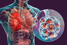
What is bronchiectasis?

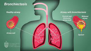
Bronchiectasis is a respiratory condition characterized by the abnormal widening and thickening of the bronchial tubes, resulting in their permanent dilation.

This disorder affects the respiratory system and can arise from various causes, including recurrent respiratory infections, cystic fibrosis, immunodeficiency disorders, and airway obstruction.

Two prominent features of bronchiectasis are chronic coughing, often accompanied by the production of excessive sputum, and recurrent respiratory infections due to the impaired clearance of mucus from the dilated bronchial tubes.

These characteristics significantly impact the quality of life of individuals with bronchiectasis and necessitate ongoing management and treatment to alleviate symptoms and prevent complications.

Causes of Bronchiectasis

  • Microorganisms: Viruses and Bacteria Bronchiectasis often arises from the repeated onslaught of respiratory infections caused by various microorganisms, such as viruses and bacteria. These pathogens invade the respiratory tract, leading to chronic inflammation and structural damage to the bronchial tubes over time.
  • Cystic Fibrosis A prevalent underlying cause of bronchiectasis is cystic fibrosis, a genetic disorder characterized by the production of thick, sticky mucus in the airways. This abnormal mucus obstructs the bronchial passages, fostering bacterial growth and chronic infection, ultimately contributing to the development of bronchiectasis.
  • Obstruction: Foreign Bodies and Tumors Obstruction of the airways, whether by foreign bodies or tumors, can significantly predispose individuals to bronchiectasis. Foreign bodies lodged in the bronchi disrupt airflow and cause localized damage, while tumors may physically block the passage of air, both impairing the normal function of the respiratory system and leading to bronchial dilation.
  • Inhalation of Corrosive Gases The inhalation of corrosive gases represents another potential cause of bronchiectasis.  Exposure to such agents directly injures the delicate lining of the airways, triggering inflammation and structural changes that contribute to the development of bronchial dilation and subsequent bronchiectasis.

Predisposing factors

  • Measles Measles, a highly contagious viral infection, can serve as a predisposing factor for bronchiectasis. The severe respiratory symptoms associated with measles, including coughing and difficulty breathing, can lead to prolonged inflammation and damage to the bronchial tubes, setting the stage for the development of bronchiectasis in susceptible individuals.
  • Broncho-pneumonia Bronchopneumonia, a type of bacterial pneumonia that affects the bronchi and surrounding lung tissue, can predispose individuals to bronchiectasis. The inflammatory response triggered by bronchopneumonia can cause scarring and structural changes in the airways, increasing the likelihood of bronchial dilation and subsequent bronchiectasis.
  • Chronic Bronchitis Chronic bronchitis, characterized by persistent inflammation of the bronchial tubes and excessive mucus production, is a common predisposing condition for bronchiectasis. The chronic irritation and damage to the airways associated with chronic bronchitis can weaken the bronchial walls, making them more susceptible to dilation and structural changes over time.
  • Tuberculosis Tuberculosis (TB), a bacterial infection primarily affecting the lungs, can predispose individuals to bronchiectasis. The destructive nature of TB on lung tissue can result in extensive scarring and fibrosis, leading to narrowing and obstruction of the bronchial passages. These structural changes increase the risk of bronchial dilation and the subsequent development of bronchiectasis in individuals with a history of TB.

Pathophysiology 

Bronchiectasis involves a complex interplay of inflammatory processes, structural changes, and impaired mucociliary clearance within the respiratory system.

The condition typically begins with an initial insult, such as recurrent respiratory infections, cystic fibrosis, or airway obstruction, which triggers inflammation in the bronchial tubes.

Chronic inflammation leads to damage of the bronchial walls and surrounding tissues, resulting in the weakening and dilation of the airways.

This bronchial dilation is accompanied by the accumulation of thickened mucus, which further obstructs airflow and creates an environment conducive to bacterial colonization and recurrent infections.

Over time, the persistent cycle of inflammation, infection, and mucus production contributes to the progressive destruction of the bronchial architecture, leading to irreversible bronchiectasis.

The dilated and distorted bronchial tubes lose their ability to effectively clear mucus and pathogens, perpetuating the cycle of inflammation and infection.

Additionally, the presence of bronchiectasis can exacerbate respiratory symptoms and impair lung function, leading to chronic cough, excessive sputum production, and recurrent respiratory infections.

Complications such as respiratory failure, pneumonia, and lung abscesses may arise in severe cases.

Signs and Symptoms

Bronchiectasis manifests with a variety of signs and symptoms, reflecting the underlying pathophysiological changes within the respiratory system.

  • Chronic Cough with Large Amounts of Purulent Sputum: One of the hallmark symptoms of bronchiectasis is a persistent cough, often accompanied by the production of copious amounts of thick, purulent sputum. This occurs due to the impaired clearance of mucus from the dilated bronchial tubes, leading to its accumulation and subsequent coughing as the body attempts to expel the excess secretion.
  • Haemoptysis: Bronchiectasis can cause episodes of haemoptysis, or coughing up blood, due to the inflammation and damage to the bronchial walls. The dilation of the bronchi increases the likelihood of blood vessels rupturing, resulting in the presence of blood in the sputum.
  • Clubbing of the Fingers: Clubbing of the fingers, characterized by the enlargement and rounding of the fingertips, is a physical manifestation commonly associated with chronic respiratory conditions, including bronchiectasis. It occurs due to chronic hypoxia and tissue inflammation, leading to changes in the structure of the nail beds and fingertips.
  • Loss of Energy, Appetite, and Weight: Bronchiectasis can lead to systemic symptoms such as fatigue, decreased appetite, and unintentional weight loss. These symptoms may result from the body’s increased metabolic demands due to chronic inflammation, as well as the energy expenditure associated with coughing and respiratory efforts.
  • Fever and Sweating at Times: Episodes of fever and sweating may occur in individuals with bronchiectasis, particularly during periods of acute exacerbation or respiratory infection. These symptoms are indicative of the body’s immune response to infection and inflammation within the respiratory tract.
  • Shortness of Breath: Patients with bronchiectasis often experience shortness of breath, especially during physical exertion or exacerbations of their condition. This symptom occurs due to the narrowing and obstruction of the airways, which impairs the normal flow of air in and out of the lungs.
  • Chest Pain: Chest pain may occur in some patients with bronchiectasis, particularly during acute exacerbations or due to underlying respiratory infections. The inflammation and irritation of the bronchial tubes can lead to discomfort or aching sensations in the chest area.
  • Wheezing: Wheezing, characterized by high-pitched whistling sounds during breathing, can occur in individuals with bronchiectasis. This symptom results from turbulent airflow through narrowed or obstructed bronchial passages.
  • Fatigue and Weakness: Chronic fatigue and weakness are common symptoms of bronchiectasis, often attributed to the body’s response to chronic inflammation, frequent coughing episodes, and respiratory infections. These symptoms can significantly impact daily activities and the overall quality of life.
  • Recurrent Respiratory Infections: Bronchiectasis predisposes patients to recurrent respiratory infections, such as bronchitis, pneumonia, and sinusitis. Frequent episodes of infection contribute to worsening symptoms and exacerbations of the underlying condition.
  • Breathlessness While Lying Flat (Orthopnea): Patients with bronchiectasis may experience difficulty breathing when lying flat, a condition known as orthopnea. This positional symptom occurs due to the redistribution of fluid within the lungs, leading to increased lung congestion and respiratory distress.
  • Cyanosis In severe cases, patients with bronchiectasis and cyanosis present with a bluish discoloration of the skin and mucous membranes. Cyanosis occurs due to inadequate oxygenation of the blood, resulting from impaired gas exchange in the lungs.

Medical Management

History

  • Respiratory Symptoms: Ask the patient about the presence and duration of symptoms such as chronic cough, excessive sputum production, shortness of breath, and recurrent respiratory infections.
  • Medical History: Ask the patient about any underlying conditions predisposing to bronchiectasis, such as cystic fibrosis, prior respiratory infections, autoimmune disorders, or a history of tuberculosis.
  • Environmental Exposures: Ask the patient about any potential exposure to environmental pollutants, occupational hazards, or inhalation of corrosive gases that could contribute to bronchiectasis.
  • Family History: Evaluate for a family history of respiratory conditions, including cystic fibrosis or bronchiectasis, which may indicate a genetic predisposition.
  • Medication History: Assess for any medications or treatments that could exacerbate or alleviate bronchiectasis symptoms, such as bronchodilators, mucolytics, or antibiotics.

Physical Examination

  • Respiratory Examination: Auscultate the lungs for abnormal breath sounds such as wheezes, crackles, or diminished breath sounds. Note any signs of respiratory distress or increased difficulty breathing.
  • Clubbing of Fingers: Inspect the fingertips for evidence of clubbing, characterized by bulbous enlargement and rounding of the nail beds.
  • Chest Examination: Palpate the chest for tenderness or deformities. Percuss the chest to assess for dullness or hyperresonance, indicative of underlying lung pathology.
  • Vital Signs: Measure vital signs, including temperature, pulse, blood pressure, and respiratory rate. Note any fever, tachycardia, or signs of hypoxia.
  • General Examination: Evaluate the patient’s general appearance, including nutritional status, hydration, and signs of systemic illness such as fatigue or weight loss.
  • Skin Examination: Look for signs of cyanosis or peripheral edema, which may indicate underlying respiratory compromise or cardiovascular involvement.
  • Neck Examination: Examine the neck for signs of lymphadenopathy or jugular venous distention, suggestive of possible infectious or inflammatory processes affecting the respiratory system.

Investigations

  • Chest X-ray: A chest X-ray will help visualize structural abnormalities in the lungs, such as bronchial dilation, thickening of bronchial walls, and areas of consolidation or atelectasis.
  • High-Resolution Computed Tomography (HRCT) Scan: HRCT imaging is considered the gold standard for diagnosing bronchiectasis. It provides detailed cross-sectional images of the lungs, allowing for precise assessment of bronchial dilatation, bronchial wall thickening, and associated lung abnormalities.
  • Pulmonary Function Tests (PFTs): PFTs assess lung function and may reveal obstructive or restrictive patterns characteristic of bronchiectasis. Common tests include spirometry, which measures lung volumes and airflow rates, and diffusion capacity testing, which evaluates gas exchange in the lungs.
  • Sputum Culture and Sensitivity: Sputum samples should be collected and analyzed to identify the presence of bacteria, viruses, or fungi causing respiratory infections. Additionally, sensitivity testing helps determine the most effective antimicrobial agents for treatment.
  • Blood Tests: Laboratory investigations such as complete blood count (CBC), inflammatory markers (e.g., C-reactive protein, erythrocyte sedimentation rate), and immunoglobulin levels may provide valuable information about the presence and severity of inflammation, infection, or immune dysfunction associated with bronchiectasis.
  • Bronchoscopy: Bronchoscopy involves inserting a flexible scope into the airways to directly visualize the bronchial tree. It allows for the assessment of the bronchial mucosa, the identification of any airway abnormalities or obstructions, and collection of samples for biopsy or culture.
  • Sputum Cytology: Sputum cytology involves examining sputum samples under a microscope to detect abnormal cells, which may indicate underlying lung cancer or other malignancies.
  • Genetic Testing: Genetic testing may be indicated in individuals with suspected cystic fibrosis or other hereditary conditions predisposing to bronchiectasis. It helps identify specific gene mutations associated with these disorders.
  • Immunological Testing: Evaluation of immune function through tests such as immunoglobulin levels, complement levels, and lymphocyte subset analysis may be warranted to assess for underlying immune deficiencies contributing to recurrent respiratory infections.
  • Lung Biopsy: In certain cases, a lung biopsy may be performed to obtain tissue samples for histological examination. This procedure is typically reserved for patients with atypical or severe presentations of bronchiectasis to rule out other pulmonary diseases or complications.

Treatment

  • Physiotherapy: Physiotherapy plays a crucial role in managing bronchiectasis by facilitating airway clearance and improving lung function. Breathing exercises, including deep breathing and controlled coughing techniques, help mobilize and expel mucus from the bronchial tubes.
  • Postural drainage involves positioning the body to facilitate the drainage of secretions from specific lung segments, enhancing clearance. Chest percussion and vibration techniques may also be utilized to loosen mucus and promote its removal.
  • High Fluid Intake: Adequate hydration is essential for individuals with bronchiectasis to help liquefy thick mucus secretions, making them easier to expectorate. Encouraging a high fluid intake, typically around 3000 milliliters per day, can aid in maintaining hydration and optimizing airway clearance.
  • Antibiotic Therapy: Antibiotics are often prescribed to treat acute respiratory infections and exacerbations of bronchiectasis. The choice of antibiotic depends on the specific pathogens identified through sputum culture and sensitivity testing.
  • Commonly used antibiotics include macrolides (e.g., azithromycin), cephalosporins, fluoroquinolones, and beta-lactam antibiotics. Long-term antibiotic therapy may also be considered in select cases to suppress chronic bacterial colonization and reduce the frequency of exacerbations.
  • Mucolytic Agents: Mucolytic agents such as dornase alfa (Pulmozyme) may be prescribed to help thin and break down thick mucus secretions, facilitating their clearance from the airways. These medications can improve respiratory symptoms and reduce the risk of respiratory infections.
  • Bronchodilators: Bronchodilators such as beta-agonists (e.g., albuterol) and anticholinergics (e.g., ipratropium bromide) may be used to alleviate bronchospasm and improve airflow in individuals with bronchiectasis. These medications help relieve symptoms such as wheezing and shortness of breath.
  • Anti-inflammatory Therapy: Inhaled corticosteroids, such as fluticasone and budesonide, may be prescribed to reduce airway inflammation and suppress the inflammatory response in bronchiectasis.
  • These medications help alleviate symptoms and may decrease the frequency of exacerbations in some patients.
  • Surgical Intervention: Surgical removal of severely damaged or infected lung tissue may be considered in cases of localized bronchiectasis that are refractory to medical management.
  • Procedures such as lobectomy (removal of a lobe of the lung) or pneumonectomy (removal of an entire lung) may be performed to improve lung function and prevent further complications.
  • Immunization: Immunization against respiratory pathogens such as influenza and pneumococcus is recommended for individuals with bronchiectasis to reduce the risk of respiratory infections and exacerbations.
  • Annual influenza vaccination and pneumococcal vaccination are particularly important to prevent serious complications in this population.

Nursing Management

Environment

  • Ensure that there is adequate ventilation in the patient’s living space to minimize exposure to indoor pollutants and allergens.
  • Implement infection control measures, including regular cleaning of surfaces and equipment, to reduce the risk of respiratory infections.
  • Create a calm and relaxing environment to reduce stress and anxiety, which can exacerbate respiratory symptoms.
  • Encourage smoking cessation and provide support for individuals who smoke or are exposed to secondhand smoke, as smoking can worsen bronchiectasis symptoms and disease progression.

Observation

  • Monitor oxygen saturation levels using a pulse oximeter regularly. Use pulse oximetry to assess respiratory function and detect hypoxemia to act as baseline data.
  • Document the frequency and severity of coughing episodes, sputum production, and any changes in respiratory symptoms.
  • Assess for signs of respiratory distress, such as increased work of breathing, use of accessory muscles, or cyanosis.
  • Track trends in vital signs, including temperature to rule out any infection, heart rate to rule out any heart failure, and respiratory rate to rule out respiratory failure, to identify patterns indicative of worsening bronchiectasis or acute exacerbations.

Psychological Care

  • Give emotional support and active listening to the patient to address fears by explaining the process of the disease, concerns, and feelings of isolation associated with living with a chronic respiratory condition.
  • Facilitate peer support groups or counseling sessions to provide opportunities for individuals with bronchiectasis to connect with others facing similar challenges.
  • Educate patients and their families about bronchiectasis, its management, and coping strategies to empower them to manage the condition.
  • Collaborate with mental health professionals to provide comprehensive psychological support and interventions tailored to the individual’s needs.
  • Develop a therapeutic nurse patient relationship to promote trust and confidence.

Hygiene

  • Educate the patients and caregivers on proper hand hygiene practices, including thorough handwashing with soap and water for at least 20 seconds.
  • Provide guidance on maintaining a clean and dust-free home environment, including regular cleaning of bedding, carpets, and upholstery.
  • Demonstrate proper technique for using respiratory hygiene measures, such as covering the mouth and nose when coughing or sneezing, and disposing of tissues promptly.
  • Encourage adherence to vaccination schedules, including annual influenza vaccination and pneumococcal vaccination, to reduce the risk of respiratory infections.

Exercises

  • Teach and encourage the performance of deep breathing exercises to promote lung expansion and improve ventilation-perfusion matching.
  • Tell the patients the proper technique for postural drainage and percussion to facilitate the mobilization and clearance of respiratory secretions.
  • Incorporate regular physical activity into the patient’s daily routine, focusing on exercises that improve cardiovascular fitness and strengthen respiratory muscles.
  • Provide education on relaxation techniques, such as guided imagery or progressive muscle relaxation, to reduce stress and tension in the chest muscles, promoting easier breathing.

Nutrition

  • Assess the patient’s nutritional status and dietary intake to identify any deficiencies or dietary modifications needed to support respiratory health.
  • Collaborate with dietitians to develop individualized meal plans that are high in nutrients and calories to meet the increased energy demands associated with chronic respiratory conditions.
  • Educate patients on the importance of staying hydrated and consuming adequate fluids to maintain optimal mucus viscosity and promote effective airway clearance.
  • Offer nutritional counseling and support to address any dietary restrictions, food preferences, or challenges related to eating with bronchiectasis.

Elimination

  • Give adequate fluid intake to prevent dehydration and maintain optimal mucus hydration for effective airway clearance.
  • Provide education on dietary fiber intake and bowel habits to promote regular bowel movements and prevent constipation, which can exacerbate respiratory symptoms.
  • Implement a toileting schedule to promote regular bowel habits and minimize the risk of fecal impaction, which can impair respiratory function.
  • Monitor urinary output and encourage frequent voiding to prevent urinary tract infections, which can increase the risk of systemic infections and exacerbate bronchiectasis symptoms.

Medicine

  • Give prescribed medications, including bronchodilators, mucolytics, and antibiotics, according to the prescribed schedule and dosage.
  • Educate the patient on the purpose, dosage, and potential side effects of each medication to promote medication adherence and optimize therapeutic outcomes.
  • Monitor for adverse reactions or medication interactions, and promptly report any concerns to the healthcare team for further evaluation and management.
  • Collaborate with the healthcare team to adjust medication regimens as needed based on the patient’s response to treatment and changes in clinical status.

Advice at Discharge

  • Give a written instructions and educational materials summarizing key self-management strategies, including medication management, airway clearance techniques, and symptom recognition.
  • Schedule follow-up appointments with healthcare providers to monitor disease progression, adjust treatment plans, and address any concerns or questions.
  • Coordinate referrals to community resources, support groups, or pulmonary rehabilitation programs to provide ongoing support and education beyond the hospital setting.
  • Encourage patients to maintain open communication with their healthcare team and seek medical attention promptly for any worsening symptoms or concerns.

Complications of Bronchiectasis

Bronchopneumonia

  • Bronchopneumonia, a type of pneumonia that affects the bronchi and surrounding lung tissue, is a common complication of bronchiectasis.
  • Chronic inflammation and mucus buildup in the dilated bronchial tubes create an ideal environment for bacterial colonization and respiratory infections, predisposing individuals with bronchiectasis to recurrent episodes of bronchopneumonia.

Cor Pulmonale

  • Cor pulmonale, also known as pulmonary heart disease, is a condition characterized by enlargement and dysfunction of the right side of the heart secondary to pulmonary hypertension and chronic lung disease.
  • In bronchiectasis, chronic inflammation and airflow obstruction lead to pulmonary hypertension, increasing the workload on the right ventricle of the heart.
  • Over time, the right ventricle may become hypertrophied and dilated, impairing its ability to pump blood effectively to the lungs, resulting in cor pulmonale.

Right-Sided Heart Failure

  • Right-sided heart failure occurs when the right ventricle of the heart is unable to pump blood effectively to the lungs, leading to fluid accumulation and congestion in the systemic venous circulation.
  • In bronchiectasis, chronic inflammation, hypoxia, and pulmonary hypertension contribute to the development of right-sided heart failure.

Respiratory Failure

  • Severe bronchiectasis can lead to respiratory failure, a condition characterized by the inability of the respiratory system to maintain adequate gas exchange.
  • Progressive airway obstruction, chronic hypoxia, and respiratory muscle fatigue contribute to respiratory failure in bronchiectasis.

Lung Abscess

  • Lung abscesses can develop in individuals with bronchiectasis as a complication of recurrent respiratory infections and impaired mucus clearance.
  • Pockets of pus form within the lung tissue, leading to localized inflammation, necrosis, and cavitation.

Hemoptysis

  • Hemoptysis, or coughing up blood, can occur as a complication of bronchiectasis due to the presence of damaged blood vessels in the dilated bronchial tubes.
  • Chronic inflammation and recurrent respiratory infections can cause erosion of blood vessels within the bronchi, leading to bleeding episodes.

Empyema

  • Empyema is a serious complication of bronchiectasis characterized by the accumulation of pus in the pleural cavity surrounding the lungs.
  • In bronchiectasis, persistent respiratory infections and lung abscesses can lead to the spread of infection to the pleural space, resulting in empyema.

Pulmonary Hypertension

  • Pulmonary hypertension, characterized by elevated blood pressure in the pulmonary arteries, can develop as a complication of advanced bronchiectasis.
  • Chronic inflammation, airway obstruction, and hypoxemia contribute to the development of pulmonary hypertension in bronchiectasis.

Lung Fibrosis

  • Lung fibrosis, or the formation of scar tissue within the lung parenchyma, can occur as a consequence of chronic inflammation and recurrent respiratory infections in bronchiectasis.
  • Progressive scarring and fibrosis impair lung function and gas exchange, leading to respiratory symptoms and reduced exercise tolerance.

Read more