Pin Worms | Causes | Signs and Symptoms | Treatment | Nursing Management

What is a pinworm?

Pinworms, also known as Enterobius vermicularis, are small parasitic worms that commonly infect the intestines of humans, particularly children.

These worms belong to the group of parasites known as helminths and are one of the most common intestinal parasites worldwide.

Pinworm infection, or enterobiasis, is highly contagious and can spread easily from person to person, especially in crowded or unsanitary conditions.

Transmission of Pinworms

Pinworms are primarily transmitted through human-to-human contact, typically by ingesting infectious pinworm eggs or through anal insertion.

These eggs are resilient and can remain viable in a moist environment for up to three weeks.

Once deposited near the anus, pinworm eggs can easily contaminate various surfaces. The sticky surface of the eggs allows them to adhere to objects such as fingernails, hands, clothing, bedding, furniture, toys, and bathroom fixtures.

Dust containing eggs can also become airborne when disturbed, potentially entering the mouth and nose through inhalation and later being swallowed.

Although pinworms do not multiply within the human body, some larvae may hatch on the anal mucosa and migrate back into the gastrointestinal tract, a process known as retroinfection.

This retroinfection can lead to a heavy parasitic load, perpetuating the infestation. Autoinfection, where the original host becomes reinfected through either the anus-to-mouth route or retroinfection, allows pinworms to persist within the same host indefinitely.

Overall, pinworm transmission occurs through a combination of contaminated surfaces, ingestion of eggs, and the unique mechanisms of retroinfection and autoinfection, contributing to the persistence of the infestation within human hosts.

Epidemiology of Pinworm Infection

Pinworm infection, caused by Enterobius vermicularis, is widespread globally and is considered the most prevalent helminth infection.

It predominantly affects children, with finger sucking and nail biting identified as behaviors that increase both the incidence and relapse rates of infection.

Transmission occurs through contamination, making pinworms common in households and among individuals in close contact.

The prevalence of pinworm infection is not influenced by gender, social class, race, or culture. However, it is more common in families with school-aged children, primary caregivers of infected children, and institutionalized children.

Pinworm eggs, deposited around the anus by female worms, can contaminate common surfaces like hands, toys, bedding, clothing, and toilet seats.

Ingesting these eggs, either directly or indirectly, through hand-to-mouth contact or inhalation, leads to infection.

After ingestion, there is an incubation period of 1 to 2 months or longer before adult gravid female worms mature in the small intestine.

Once mature, female worms migrate to the colon and lay eggs around the anus at night, often while hosts are asleep. Infected individuals can transmit the parasite to others as long as female pinworms continue depositing eggs on the perianal skin.

Reinfection is possible through self-inoculation or exposure to eggs from another person. The primary symptom of pinworm infection, known as enterobiasis, is itching in the anal area.

Overall, pinworm infection is highly prevalent, especially among children, and transmission occurs through various routes of contamination and ingestion of eggs.
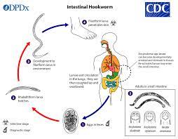
Life Cycle

The entire life cycle of pinworms, from egg to adult, occurs within the human gastrointestinal tract of a single host. The life cycle commences with the ingestion of pinworm eggs, which hatch in the duodenum.

The newly hatched larvae, measuring approximately 140 to 150 micrometers in size, rapidly grow as they migrate through the small intestine towards the colon. During this migration, the larvae undergo two molts and mature into adult pinworms.

Female pinworms typically survive for 5 to 13 weeks, while males live approximately 7 weeks. Mating between male and female pinworms occurs in the ileum, after which the male pinworms usually die and are expelled from the host via stool.

Gravid female pinworms settle in various regions of the large intestine, including the ileum, cecum, appendix, and ascending colon, where they attach themselves to the intestinal mucosa and feed on colonic contents. The gravid females become filled with eggs, with estimates ranging from about 11,000 to 16,000 eggs per female.

Egg-laying begins approximately five weeks after the initial ingestion of pinworm eggs by the human host. Gravid female pinworms migrate towards the rectum at a rate of 12 to 14 centimeters per hour.

Upon reaching the anus, the female pinworms emerge and deposit eggs onto the skin surrounding the anus through various means, including contracting and expelling eggs, disintegrating upon death, or rupturing due to host scratching.

Following egg deposition, the female pinworm becomes opaque and dies. The emergence of the female from the anus serves to obtain oxygen necessary for egg maturation.

Causes of Pinworm 

  • Ingestion of Pinworm Eggs: The primary cause of pinworm infection is the ingestion of pinworm eggs. These eggs are commonly found in contaminated food, water, or on surfaces touched by infected individuals.
  • Direct Transmission: Pinworms are highly contagious and can spread directly from person to person through contact with contaminated hands, objects, or surfaces.
  • Indirect Transmission: Pinworm eggs can also be transmitted indirectly through the ingestion or inhalation of airborne dust contaminated with eggs. This can occur when eggs are disturbed by surfaces and become airborne.
  • Poor Hygiene Practices: Inadequate hand hygiene, such as not washing hands properly after using the toilet or before handling food, can contribute to the spread of pinworm infection.
  • Close Contact: Pinworm infection is more common in settings where individuals live in close quarters, such as households, schools, daycares, and institutional settings, due to the ease of transmission in such environments.
  • Fomite Transmission: Pinworm eggs can survive for extended periods on surfaces such as bedding, clothing, toys, and furniture. Contact with contaminated foods can lead to the transmission of the infection.
  • Autoinfection: In some cases, individuals can become reinfected with pinworms by ingesting eggs that were previously expelled from their own bodies. This autoinfection can occur through activities such as scratching the anal area and then touching the mouth without proper hand hygiene.

Signs and Symptoms

  • Anal Itching: The most common symptom of pinworm infection is intense itching around the anus, particularly at night. This itching is caused by the movement of female pinworms as they lay eggs around the anal area.
  • Restlessness: Infected individuals may experience restlessness, especially at night, due to discomfort caused by anal itching.
  • Difficulty Sleeping: The itching sensation around the anus can disrupt sleep patterns, leading to difficulty falling asleep or staying asleep throughout the night.
  • Irritability: Chronic itching and disrupted sleep can contribute to irritability and mood changes, particularly in children.
  • Abdominal Pain: Some individuals may experience mild abdominal pain or discomfort, although this symptom is less common than anal itching.
  • Loss of Appetite: In severe cases of pinworm infection, loss of appetite may occur, possibly due to discomfort or gastrointestinal disturbances.
  • Secondary Infections: Excessive scratching of the anal area can lead to skin irritation, inflammation, and secondary bacterial infections.
  • Vaginal Itching: In females, pinworms may migrate to the vaginal area, causing itching and discomfort.
  • Bedwetting: Pinworm infection may contribute to bedwetting, particularly in children, due to disrupted sleep patterns and discomfort.
  • Weight Loss: Severe and prolonged pinworm infections may result in weight loss, although this is rare and usually associated with complications such as malnutrition.

Medical Management

Patient History

  • Ask about any symptoms such as anal itching, particularly at night, restlessness, difficulty sleeping, abdominal pain, and irritability.
  • Ask about recent exposures to potential sources of infection, such as contact with individuals known to have pinworm infection or exposure to contaminated surfaces or environments.
  • Explore any history of previous pinworm infections or treatments.

Physical Examination

  • Inspect the perianal area for signs of inflammation, redness, or excoriation due to scratching.
  • Look for visible evidence of pinworms or their eggs around the anus, such as tiny white threads or specks.
  • Check for signs of secondary bacterial skin infections resulting from excessive scratching.
  • Assess for any abdominal tenderness or discomfort that may indicate a more severe infection or complications.

Additional Considerations

  • In pediatric patients, ask about symptoms such as irritability, difficulty concentrating, or changes in behavior that may indicate pinworm infection.
  • Consider performing a scotch tape test or cellophane tape test to collect samples from the perianal area for microscopic examination to confirm the presence of pinworm eggs.
  • Discuss the possibility of household or close contacts also undergoing screening and treatment if warranted based on history and examination findings.
  • Rule out other potential causes of anal itching or gastrointestinal symptoms, such as other parasitic infections, dermatological conditions, or gastrointestinal disorders.

Investigations 

  • Scotch Tape Test: This simple and non-invasive test involves pressing a piece of clear adhesive tape against the skin around the anus in the morning before bathing or using the toilet. The tape is then transferred to a glass slide and examined under a microscope for the presence of pinworm eggs.
  • Stool Examination: In some cases, a stool sample may be collected from the patient and examined for the presence of pinworm eggs or adult worms. This can be particularly useful if the infection is suspected to be more severe or if other gastrointestinal symptoms are present.
  • Perianal Swab: A cotton swab may be used to collect samples from the perianal area for microscopic examination to detect pinworm eggs or evidence of infection.
  • Serological Tests: While less commonly used, serological tests may be performed to detect antibodies against pinworm antigens in blood samples. These tests are not routinely recommended for diagnosis but may be useful in certain situations, such as when other methods are inconclusive or to monitor response to treatment.
  • Diagnostic Imaging: In rare cases where complications of pinworm infection are suspected, such as intestinal obstruction or appendicitis, diagnostic imaging studies such as abdominal ultrasound or computed tomography (CT) scan may be performed to assess the extent of the infection and associated complications.

Treatment 

  • The many primary drugs used for treating pinworm infection include mebendazole, pyrantel pamoate, and albendazole.
  • Treatment typically involves administering a single dose of the medication initially, followed by a second dose two weeks later. This two-dose regimen helps to ensure the elimination of both adult worms and any newly hatched larvae.
  • It’s important to note that these medications may not reliably kill pinworm eggs, hence the need for the second dose to prevent re-infection by adult worms hatching from any surviving eggs.
  • When considering treatment for children under 2 years of age, health practitioners and parents should carefully evaluate the potential risks and benefits of these drugs.
  • In cases of repeated infections, the same treatment method as for the initial infection should be followed.
  • For households with multiple infected individuals or instances of recurrent symptomatic infections, it is advisable to treat all household members simultaneously to prevent ongoing transmission.
  • In institutional settings, mass and simultaneous treatment of all individuals, repeated after two weeks, can be an effective strategy to control the spread of pinworm infection.

Nursing Management 

Aims

  • Alleviate the symptoms and discomfort associated with pinworm infection.
  • Prevent the transmission of pinworms to others.
  • Educate patients and caregivers about preventive measures and treatment adherence.
  • Promote the overall health and well-being of patients affected by pinworm infection.

Environment

  • Ensure cleanliness and hygiene in the living environment, including regular cleaning of surfaces and bedding to remove pinworm eggs.
  • Encourage the patient to understand the importance of frequent handwashing with soap and water, especially after using the toilet and before handling food.
  • Promote proper disposal of contaminated items, such as underwear and bedding, to prevent re-infection.
  • Educate caregivers about the importance of maintaining a hygienic environment to reduce the risk of pinworm transmission.

Observation

  • Monitor patients for symptoms of pinworm infection, such as anal itching, restlessness, and difficulty sleeping.
  • Assess the perianal area for signs of irritation, redness, or excoriation due to scratching.
  • Observe for behavioral changes, such as irritability or changes in appetite, which may indicate discomfort associated with the infection.
  • Document any changes in symptoms or treatment responses to guide further management and intervention.

Psychological Care

  • Provide emotional support and reassurance to patients experiencing discomfort or embarrassment due to a pinworm infection.
  • Offer education and counseling to alleviate anxiety and concerns about transmission to others.
  • Encourage open communication and discussion of feelings related to the infection to promote psychological well-being.
  • Collaborate with other healthcare professionals, such as psychologists or social workers, as needed for additional support and intervention.

Hygiene

  • Educate patients and caregivers about proper handwashing techniques and the importance of hand hygiene in preventing pinworm transmission.
  • Instruct patients to keep fingernails short and clean to reduce the risk of transferring pinworm eggs from the hands to the mouth.
  • Emphasize the importance of regular bathing and changing of underwear to maintain personal hygiene and reduce the spread of infection.
  • Provide guidance on laundering clothing, bedding, and towels in hot water to kill any pinworm eggs and prevent re-infection.

Exercises

  • Encourage regular physical activity and exercise to promote overall health and well-being.
  • Discuss the benefits of outdoor play and recreational activities in reducing sedentary behavior and promoting immune function.
  • Provide age-appropriate exercise recommendations and activities for children to engage in both indoors and outdoors.
  • Collaborate with physical therapists or exercise specialists as needed to develop tailored exercise programs for patients with specific needs or limitations.

Nutrition

  • Offer guidance on maintaining a balanced diet rich in fruits, vegetables, whole grains, and lean proteins to support immune function and overall health.
  • Provide information on foods high in fiber to promote regular bowel movements and aid in the elimination of pinworms from the digestive tract.
  • Address any concerns or questions related to dietary restrictions or recommendations during pinworm infection and treatment.
  • Collaborate with dietitians or nutritionists as needed to develop personalized nutrition plans for patients with specific dietary needs or preferences.

Elimination

  • Educate patients and caregivers about the importance of proper hygiene practices after using the toilet, including thorough wiping and handwashing.
  • Instruct patients to avoid scratching the anal area to prevent irritation and the potential spread of pinworm eggs.
  • Provide guidance on the proper disposal of feces to minimize the risk of contaminating the environment with pinworm eggs.
  • Monitor bowel habits and stool consistency to assess treatment response and ensure effective elimination of pinworms from the digestive tract.

Medicine

  • Administer prescribed medications, such as mebendazole or albendazole, according to the recommended dosage and schedule.
  • Educate patients and caregivers about the purpose, benefits, and potential side effects of the prescribed medications.
  • Encourage adherence to the full course of treatment, including any follow-up doses or instructions for re-treatment.
  • Monitor for adverse reactions or complications related to medication therapy, and report any concerns to the healthcare provider for further evaluation.

Advice at Discharge

  • Provide information and resources to patients and caregivers about community support services or assistance programs available for individuals facing financial or social disadvantages.
  • Offer guidance on accessing affordable healthcare services, including clinics, community health centers, and free or low-cost medication programs.
  • Advocate for patients’ rights and access to quality healthcare, including timely diagnosis and treatment of pinworm infection.
  • Collaborate with social workers or case managers to address social determinants of health that may impact patients’ ability to manage pinworm infection effectively.

Prevention 

  • Hand Hygiene: Washing hands thoroughly with soap and warm water after using the toilet, changing diapers, and before handling food is the most effective way to prevent pinworm infection.
  • Daily Bathing: Infected individuals should bathe every morning to remove a large number of pinworm eggs on the skin. Showering is preferable to taking a bath to avoid contaminating bath water with eggs. Infected individuals should not share baths with others during their infection period.
  • Nail Care: Regularly cutting fingernails and avoiding nail biting can help prevent the transfer of pinworm eggs from the hands to the mouth. Scratching around the anus should also be avoided.
  • Clothing and Bed Linens: Changing underclothes and bed linens frequently, particularly in the morning, can reduce the risk of transmitting eggs into the environment and the chance of reinfection. These items should be laundered in hot water and dried in a hot dryer to kill any eggs present.
  • Environmental Hygiene: Avoid shaking clothing and bedding to prevent the spread of pinworm eggs. Instead, carefully place them in a washer for laundering. Additionally, maintaining cleanliness in the living environment, including regular cleaning of surfaces, can help prevent the spread of infection.
  • Education: Teach children the importance of handwashing to prevent infection. In institutional settings such as daycares, schools, and healthcare facilities, control of pinworm can be challenging, but mass drug administration during outbreaks can be effective.

Complications of Pinworm Infection

  • Secondary Bacterial Infections: Excessive scratching of the perianal area due to itching caused by pinworm infection can lead to skin irritation and breakdown, increasing the risk of bacterial infections such as cellulitis or impetigo.
  • Sleep Disturbance: Persistent itching, particularly at night when female pinworms lay their eggs, can disrupt sleep patterns and lead to insomnia or poor quality sleep, impacting overall well-being and daily functioning.
  • Psychological Distress: Chronic or recurrent pinworm infection can cause psychological distress, including anxiety, embarrassment, and social isolation, especially in children and adolescents.
  • Complications in Rare Cases: Although rare, severe complications of pinworm infection may include appendicitis, urinary tract infections, or vaginal infections (in females) due to the migration of pinworms to other parts of the body.
  • Chronic Infection: If left untreated or inadequately managed, pinworm infection can become chronic, leading to persistent symptoms and complications over time.
  • Recurrent Infections: Despite treatment, some individuals may experience recurrent pinworm infections, especially in settings where hygiene measures are inadequate or reinfection occurs due to close contact with infected individuals.
  • Impact on Quality of Life: Pinworm infection and its associated symptoms, such as anal itching and sleep disturbances, can significantly impact an individual’s quality of life, leading to discomfort, frustration, and reduced overall well-being.
  • Transmission to Others: Failure to control pinworm infection can result in transmission to other household members, caregivers, or close contacts, leading to outbreaks within communities or institutions.

Read more

Hook Worm Disease | Causes | Pathophysiology | Signs and Symptoms | Treatment | Nursing Management

What is a hookworm? 

Hookworm disease, also known as hookworm infection or ancylostomiasis, is a parasitic infection caused by hookworms. There are two main species of hookworms that infect humans: Ancylostoma duodenale and Necator americanus. These parasites primarily affect the small intestine of humans.

Hookworm infection poses a significant health burden, particularly in the developing regions of the tropics and subtropics, where it is a major cause of maternal and child morbidity. Susceptible children, when infected with hookworms, can experience intellectual, cognitive, and growth impairments. Additionally, maternal infection during pregnancy can lead to intrauterine growth retardation, prematurity, and low birth weight in newborns. While hookworm infection itself is seldom fatal, it can result in significant anemia, particularly in individuals with heavy worm burdens.

Epidemiology

It is currently estimated that between 576 and 740 million people worldwide are harboring hookworm infections. Among this population, approximately 80 million individuals suffer from severe manifestations of the disease.

Causes of Hookworm Infection

Species of Hookworms: Two species of hookworms commonly infect humans: Ancylostoma duodenale and Necator americanus.

Prevalence

  • Necator americanus predominates in the Americas, Sub-Saharan Africa, Southeast Asia, China, and Indonesia.
  • Ancylostoma duodenale predominates in the Middle East, North Africa, India, and previously in southern Europe.

Disease Presentation

  • Hookworm infection leads to ankylostomiasis, also known as anchylostomiasis or helminthiasis.
  • The disease occurs when hookworms, present in large numbers, feed voraciously on blood from the host’s intestinal walls, resulting in iron deficiency anemia.

Morphology of Ancylostoma duodenale Worms

  • Ancylostoma duodenale worms appear greyish-white or pinkish, with the head slightly bent in relation to the rest of the body.
  • They have well-developed mouths equipped with two pairs of teeth.
  • Males typically measure about one centimeter in length and 0.5 millimeters in width, while females are often longer and more robust in size.

The life cycle of Hookworms

  • Eggs in the Environment: The life cycle begins when adult female hookworms in the small intestine of the host produce eggs. These eggs are passed out through the host’s feces into the environment.
  • Larval Development: In favorable conditions of warmth, moisture, and oxygen, the eggs hatch into larvae within the soil.
  • Infective Larvae: The larvae molt twice to become infective larvae (L3 stage), which can penetrate the skin of the host, usually through bare feet, during contact with contaminated soil.
  • Migration to the Small Intestine: Once penetrated, the larvae migrate through the bloodstream to the lungs, ascend the respiratory tract, are swallowed, and eventually reach the small intestine.
  • Maturation into Adults: In the small intestine, the larvae mature into adult hookworms. They attach to the intestinal mucosa and begin feeding on the host’s blood.
  • Egg Production: Adult female hookworms produce eggs, which are passed out in the host’s feces, completing the life cycle.

Life Cycle of N. americanus and A. duodenale in the Environment

  • Eggs of N. americanus and A. duodenale are commonly found in warm, moist soil, where they undergo development until they hatch into first-stage larvae.
  • These newly hatched larvae enter a feeding, non-infective stage known as the rhabditoform stage. During this stage, they feed on soil microbes.
  • After a period of feeding, the larvae molt into second-stage larvae, which also remain in the rhabditoform stage. They continue to feed for approximately 7 days.
  • Following this feeding period, the larvae undergo another molt, transitioning into the third-stage larvae.
  • The third-stage larvae, known as the filariform stage, are the non-feeding infective form of the parasite. They are capable of penetrating the skin of the host to initiate infection.

Life Cycle In Man

  • Larvae enter the lungs through the pulmonary capillaries and break out into the alveoli.
  • They travel up the trachea and are coughed up and swallowed by the host.
  • After ingestion, larvae are found in the small intestine, where they molt into the adult worm stage.
  • The entire process, from skin penetration to adult development, takes about 5–9 weeks.
  • Female adult worms release eggs (N. Americanus: about 9,000–10,000 eggs/day; A. duodenale: 25,000–30,000 eggs per day), which are passed in the feces of the human host.
  • These eggs hatch in the environment within several days, initiating a new cycle.

The incubation period

The incubation period of hookworm infection can range from a few weeks to several months, and its duration largely depends on the severity of the infestation, determined by the number of hookworm parasites present in the individual.

In cases where the infection involves a high burden of parasites, symptoms may manifest more rapidly, leading to a shorter incubation period. Conversely, patients with lower levels of infestation tend to experience a more prolonged incubation period before symptoms become apparent.

Pathophysiology

Hookworm infection is typically considered asymptomatic, yet it poses significant danger due to its “silent and insidious” nature. While patients may initially experience general symptoms soon after infection, such as ground-itch, a common allergic reaction at the site of larval entry, particularly in N. americanus infections, there may be additional manifestations as the infection progresses.

  • As the larvae break into the alveoli and ascend the trachea, symptoms like coughing and pneumonitis may arise.
  • Upon reaching the small intestine and beginning maturation, infected individuals commonly suffer from diarrhea and gastrointestinal discomfort.

However, the true insidiousness of hookworm infection lies in chronic, heavy-intensity cases. Major morbidities stem from intestinal blood loss, iron deficiency anemia, and protein malnutrition. This results primarily from adult hookworms ingesting blood, rupturing red blood cells, and degrading hemoglobin in the host.

  • Chronic blood loss can lead to physical manifestations like facial and peripheral edema, as well as eosinophilia and pica due to iron deficiency anemia.
  • It’s widely recognized that children with chronic hookworm infection may experience growth retardation, intellectual impairment, and cognitive deficits due to the long-term effects of the parasite on their health.

Signs and Symptoms

  • Gastrointestinal Inflammation: Symptoms in patients often arise due to inflammation in the gut caused by the feeding activity of hookworms.
  • Nausea: Patients may experience feelings of nausea, particularly as a result of gut irritation.
  • Abdominal Pain and Intermittent Diarrhea: Common symptoms include abdominal pain and sporadic episodes of diarrhea, which can be linked to the inflammatory response and damage caused by the parasites in the intestines.
  • Progressive Anemia: In cases of prolonged infection, progressive anemia may develop due to chronic blood loss from the intestinal mucosa, leading to symptoms such as fatigue, weakness, and pallor.
  • Capricious Appetite and Pica: Affected people may exhibit changes in appetite, ranging from unpredictability to the development of pica, a condition characterized by cravings for non-food items such as dirt or clay, often associated with nutritional deficiencies.
  • Gastrointestinal Disturbances: Symptoms may include obstinate constipation followed by episodes of diarrhea, reflecting the disruptive effect of the parasites on normal bowel function.
  • Cardiovascular Symptoms: Palpitations, a thready pulse, and coldness of the skin may occur, reflecting the impact of anemia on cardiovascular function.
  • General Weakness and Fatigue: Individuals may experience overall fatigue and weakness due to the combined effects of anemia and systemic inflammation.
  • Shortness of Breath: Anemia and reduced oxygen-carrying capacity in the blood may lead to symptoms of dyspnea or shortness of breath.
  • Severe Complications: In severe cases, the infection may lead to complications such as dysentery, hemorrhages, and edema, potentially resulting in a fatal outcome if left untreated.

Medical Management

History

  • Travel History: A nurse should ask about any recent travel to regions where hookworm infection is prevalent, particularly tropical and subtropical areas with poor sanitation.
  • Exposure to Contaminated Environments: Ask about exposure to soil or water sources potentially contaminated with hookworm larvae.
  • Symptomatology: Make sure that a nurse evaluates for symptoms such as abdominal pain, diarrhea, nausea, fatigue, weakness, shortness of breath, and changes in appetite.
  • Medical History: Obtain information about any previous episodes of gastrointestinal disorders, anemia, or unexplained fatigue.
  • Occupational and Living Conditions: Assess living conditions, including sanitation facilities, and inquire about occupations involving soil exposure.

Physical Examination

  • General Appearance: Assess for signs of pallor, fatigue, and overall weakness, which may indicate anemia and the systemic effects of the infection.
  • Skin Examination: Look for signs of ground-itch or dermatitis at potential sites of larval penetration, such as the feet.
  • Abdominal Examination: Palpate the abdomen for tenderness, distention, or masses, and assess bowel sounds. Abdominal pain and tenderness may suggest gastrointestinal inflammation.
  • Cardiovascular Examination: Check for any signs of tachycardia, palpitations, or hypotension, which may indicate anemia-related cardiovascular compromise.
  • Respiratory Examination: Evaluate for signs of respiratory distress, cough, or abnormal breath sounds, which could indicate pulmonary involvement if larvae have migrated to the lungs.
  • Nutritional Assessment: Look for signs of malnutrition, including muscle wasting, edema, and skin changes such as dryness or scaling.
  • Neurological Examination: Assess cognitive function and look for signs of neurological deficits, particularly in children, as chronic hookworm infection can affect intellectual development.

Investigations

  • Stool Examination: The microscopic examination of stool samples to check for the presence of hookworm eggs is the primary diagnostic test for confirming hookworm infection. Eggs can be detected using various techniques, such as direct smears, concentration methods (e.g., sedimentation or flotation), or fecal immunoassays.
  • Complete Blood Count (CBC): A CBC will reveal signs of anemia, such as low hemoglobin and hematocrit levels, as well as microcytic or hypochromic red blood cells, which are suggestive of iron deficiency anemia commonly associated with hookworm infection.
  • Peripheral Blood Smear: Examination of a peripheral blood smear will reveal characteristic changes associated with anemia, such as hypochromic red blood cells and increased numbers of reticulocytes.
  • Serum Iron Studies: Measurement of serum iron, ferritin, and total iron-binding capacity (TIBC) can help assess iron status and confirm iron deficiency anemia.
  • Stool Occult Blood Test: This test detects the presence of blood in stool samples, which may indicate intestinal bleeding caused by hookworm infection.
  • Serological Tests: Serological assays, such as enzyme-linked immunosorbent assays (ELISA), can detect antibodies against hookworm antigens and may be useful for confirming infection, especially in cases where stool examination is inconclusive.
  • Imaging Studies: In severe cases or when complications such as intestinal obstruction or perforation are suspected, imaging modalities such as abdominal ultrasound or computed tomography (CT) scans may be employed to assess the extent of gastrointestinal involvement.
  • Endoscopic Evaluation: In cases of suspected gastrointestinal bleeding or inflammatory bowel disease-like symptoms, upper gastrointestinal endoscopy or colonoscopy may be performed to visualize the mucosa and obtain biopsy samples for histological examination.

Treatment

  • Albendazole Efficacy: Albendazole is effective against hookworm infection, targeting both the intestinal stage and the larval stage while the parasite is still migrating under the skin. It works by disrupting the function of the parasite’s β-tubulin protein, inhibiting microtubule polymerization, and ultimately killing the adult worms.
  • Iron Supplementation: In cases of anemia resulting from hookworm infection, iron supplementation is very important to alleviate the symptoms of iron deficiency anemia. However, as red blood cell levels are restored, deficiencies in other essential nutrients such as folic acid or vitamin B12 may arise. Then, supplementation with these nutrients may also be necessary to prevent further complications.
  • Benzimidazoles (BZAs): The primary treatment for hookworm infection involves the use of benzimidazole anthelmintic medications, specifically albendazole and mebendazole. These drugs work by binding to the nematode’s β-tubulin, disrupting microtubule polymerization, and ultimately killing the adult worms.
  • Alternative Treatments: In certain circumstances, alternative medications such as levamisole and pyrantel pamoate may be used. These drugs also target the nematode’s nervous system, leading to paralysis and the expulsion of the worms from the intestines.
  • Treatment Duration: The duration of treatment typically ranges from 1 to 3 days, depending on the severity of the infection and the specific medication used. Repeat doses may be necessary in cases of persistent or recurrent infections.
  • Mass Drug Administration (MDA): In regions where hookworm infection is endemic, mass drug administration programs may be implemented to control the spread of the disease. These programs involve the distribution of anthelmintic medications to entire communities at regular intervals to reduce the prevalence of infection.
  • Monitoring and Follow-Up: After treatment, patients should be monitored for resolution of symptoms and signs of anemia. Follow-up stool examinations may also be performed to confirm clearance of the infection. Additionally, efforts to improve sanitation and hygiene practices are essential for preventing reinfection and controlling the spread of hookworm infection within communities.

Nursing Diagnosis

  1. Impaired Growth and Development
  2. Inadequate Nutrition
  3. Diarrhea
  4. Fatigue
  5. Risk for Fluid Volume Deficit
  6. Risk for Infection
  7. Lack of Knowledge
  8. Ineffective Breathing Pattern

Nursing Management

Aims

  1. Alleviate Symptoms: The primary aim is to alleviate symptoms associated with hookworm infection, including abdominal pain, diarrhea, anemia, and fatigue, through appropriate nursing interventions and supportive care.
  2. Prevent Transmission: Nurses strive to prevent the transmission of hookworm disease by educating patients and communities about proper sanitation and hygiene practices, including wearing shoes in endemic areas and avoiding contact with contaminated soil or feces.
  3. Provide Education: Nurses play a crucial role in providing education to patients and caregivers about the transmission, symptoms, and treatment of hookworm disease, as well as preventive measures to minimize the risk of reinfection and complications.
  4. Supportive Care: Nurses provide supportive care to individuals affected by hookworm disease, including nutritional support to address deficiencies caused by chronic blood loss, psychological support to cope with the emotional impact of the disease, and monitoring for complications or adverse effects of treatment.

Environment

  • A nurse should make sure there is a clean and hygienic environment to prevent further infection. Maintain a comfortable and quiet atmosphere to promote rest and a quick recovery.
  • Make sure to implement infection control measures, such as hand hygiene and environmental sanitation. Then provide adequate ventilation and temperature control to enhance patient comfort.

Observation

  • Monitor vital signs regularly to assess for signs of dehydration or infection. Observe for signs of fatigue, weakness, and respiratory distress.
  • Monitor stool frequency, consistency, and color to evaluate diarrhea and assess for improvement. Document intake and output to monitor fluid balance and detect signs of dehydration.

Psychological Care

  • Provide emotional support to the patient and reassurance to alleviate anxiety and stress. Encourage open communication with the patient and address any concerns or fears the patient may have.
  • Give education to the patient about the infection, its treatment, and the importance of adherence to medication and hygiene practices. Collaborate with the healthcare team to address any psychosocial issues or mental health concerns.

Hygiene

  • Teach proper hand hygiene techniques to prevent the spread of infection to other patients in the ward. Assist with personal hygiene activities, such as bathing and grooming, as needed.
  • Ensure clean and sanitized bedding and clothing to minimize the risk of re-infection. Educate the patient and family members on environmental hygiene practices to reduce exposure to contaminated soil or water.

Exercises

  • Encourage light physical activity, such as walking or stretching, to promote circulation and prevent muscle weakness. Provide guidance on breathing exercises to improve lung function and alleviate respiratory symptoms.
  • Collaborate with physical therapy for customized exercise programs tailored to the patient’s condition and capabilities. Monitor the patient during exercise sessions and adjust activities as needed based on their tolerance and energy levels.

Nutrition

  • Make sure to check the dietary intake and nutritional status to identify deficiencies and develop individualized meal plans. Offer small, frequent meals and snacks to maintain energy levels and support healing.
  • Provide education on foods rich in iron, vitamins, and minerals to promote recovery from anemia and enhance immune function. Collaborate with a dietitian to address specific nutritional needs and monitor weight changes.

Elimination

  • Monitor bowel habits and provide interventions to manage diarrhea and promote regular bowel movements. Offer dietary modifications, such as increasing fiber intake or avoiding irritating foods, to regulate bowel function.
  • Administer prescribed medications, such as antidiarrheals or stool softeners, as ordered by the healthcare provider. Educate the patient on proper toilet hygiene and encourage frequent handwashing to prevent the spread of infection.

Medication

  • Administer anthelmintic medications, such as albendazole or mebendazole, as prescribed by the healthcare provider. Monitor for adverse reactions to medications and report any concerns to the healthcare team.
  • Educate the patient on the purpose, dosage, and potential side effects of prescribed medications. Ensure compliance with the medication regimen and provide reminders as needed to promote treatment effectiveness.

Advice at Discharge

  • Emphasize the importance of completing the full course of prescribed medications to ensure the eradication of the infection. Provide instructions on proper hygiene practices, including handwashing and avoiding contact with contaminated soil.
  • Encourage follow-up appointments with healthcare providers for monitoring and assessment of treatment effectiveness. Offer resources and support services, such as community health clinics or support groups, for ongoing care and assistance with managing the infection.

Prevention 

Sanitation Practices

  • Avoid defecating in open areas, and use designated latrines or toilets to dispose of waste properly.
  • Refrain from using human excrement, raw sewage, or untreated “night soil” as fertilizer in agriculture, as these can serve as breeding grounds for hookworm larvae.

Deworming

  • Implement regular deworming programs in communities, especially in areas where hookworm infection is prevalent.
  • Deworming medications can help eliminate existing infections and prevent re-infections.

Hand Hygiene

  • Promote proper handwashing practices, particularly after using the toilet, before preparing or consuming food, and after handling soil or waste.
  • Encourage the use of soap and clean water for thorough handwashing, as this helps remove hookworm larvae and prevents transmission.

Footwear Use

  • Wear shoes or protective footwear, especially in areas where soil contamination with hookworm larvae is likely, such as sandy or loamy soil.
  • Cover feet properly to minimize skin contact with contaminated soil and reduce the risk of larval penetration.

Public health education

  • Conduct public health education campaigns to raise awareness about the transmission and prevention of hookworm infections.
  • Provide information about the importance of sanitation, hygiene, and deworming in preventing infection and improving community health.

Safe Agricultural Practices

  • Educate farmers and agricultural workers about the risks associated with using human waste as fertilizer and promote the use of safer alternatives.
  • Encourage the proper composting and treatment of organic waste to eliminate pathogens, including hookworm larvae, before using it in agriculture.

Environmental Management

  • Implement measures to improve environmental sanitation, such as proper waste disposal, sewage treatment, and soil management practices.
  • Drain stagnant water sources and minimize conditions conducive to the survival and proliferation of hookworm larvae in the environment.

Community Engagement

  • Engage community members in efforts to prevent hookworm infection, including participation in deworming campaigns, sanitation improvement initiatives, and hygiene promotion activities.
  • Foster community ownership and collaboration to sustain long-term prevention efforts and reduce the burden of hookworm infection in endemic areas.

Complications

  • Anemia: Chronic hookworm infection can lead to iron deficiency anemia due to the loss of blood caused by the parasites feeding on the host’s intestinal mucosa.
  • Malnutrition: Prolonged hookworm infection can result in malabsorption of nutrients, leading to protein-energy malnutrition and deficiencies in essential vitamins and minerals.
  • Growth Retardation: Children with chronic hookworm infection may experience stunted growth and delayed development due to the nutritional deficiencies and chronic inflammation associated with the infection.
  • Cognitive Impairment: Severe hookworm infection, particularly in children, can impair cognitive function and intellectual development, leading to learning difficulties and poor academic performance.
  • Intestinal Complications: Inflammatory reactions in the intestines caused by hookworms can result in symptoms such as abdominal pain, diarrhea, and gastrointestinal bleeding.
  • Respiratory Symptoms: Migration of hookworm larvae through the lungs can cause respiratory symptoms such as cough, wheezing, and shortness of breath.
  • Skin Disorders: Ground itch, a common symptom of hookworm infection, can lead to dermatitis, itching, and skin irritation at the site of larval penetration.
  • Secondary Infections: A persistent hookworm infection can weaken the immune system, increasing the risk of secondary bacterial, fungal, or parasitic infections.
  • Pregnancy Complications: Hookworm infection during pregnancy can lead to complications such as intrauterine growth restriction, preterm birth, and low birth weight in newborns.
  • Cardiovascular Effects: Severe anemia resulting from hookworm infection can strain the cardiovascular system, leading to symptoms such as palpitations, fainting, and chest pain.
  • Mental Health Issues: Chronic illness and its associated symptoms can have psychological effects, including depression, anxiety, and reduced quality of life.
  • Fatalities: Although rare, severe complications of hookworm infection, such as massive gastrointestinal bleeding or severe anemia, can lead to fatalities if left untreated.

Read more